. 2012 Apr;97(1):105-16.
doi: 10.1016/j.exer.2012.02.004. Epub 2012 Mar 9.

Characterization of multiple light damage paradigms reveals regional differences in photoreceptor loss

Affiliations

Characterization of multiple light damage paradigms reveals regional differences in photoreceptor loss

Jennifer L Thomas et al. Exp Eye Res. 2012 Apr.

Abstract

Zebrafish provide an attractive model to study the retinal response to photoreceptor apoptosis due to its remarkable ability to spontaneously regenerate retinal neurons following damage. There are currently two widely-used light-induced retinal degeneration models to damage photoreceptors in the adult zebrafish. One model uses constant bright light, whereas the other uses a short exposure to extremely intense ultraviolet light. Although both models are currently used, it is unclear whether they differ in regard to the extent of photoreceptor damage or the subsequent regeneration response. Here we report a thorough analysis of the photoreceptor damage and subsequent proliferation response elicited by each individual treatment, as well as by the concomitant use of both treatments. We show a differential loss of rod and cone photoreceptors with each treatment. Additionally, we show that the extent of proliferation observed in the retina directly correlates with the severity of photoreceptor loss. We also demonstrate that both the ventral and posterior regions of the retina are partially protected from light damage. Finally, we show that combining a short ultraviolet exposure followed by a constant bright light treatment largely eliminates the neuroprotected regions, resulting in widespread loss of rod and cone photoreceptors and a robust regenerative response throughout the retina.


Figures

Figure 1
Figure 1. Rod photoreceptor loss following various light damage paradigms
EGFP-positive rod photoreceptors in the dorsal (A, B, E, F) and ventral (C, D, G, H) halves of the retina from adult Tg(nrd:egfp)/alb zebrafish. A. Untreated (0 hr) dorsal retina with EGFP-positive somas in the outer nuclear layer (ONL), rod inner segments (RIS), and rod outer segments (ROS). B. Dorsal retina following a 48 hour exposure to constant bright light (48 hLt), with fewer EGFP-positive somas in the ONL, and truncated RIS and ROS. C. 0 hr ventral retina with undamaged ONL, RIS, and ROS. D. 48 hLt ventral retina, with fewer ONL nuclei, but intact RIS and ROS. E. Dorsal retina 48 hours after a 30 minute exposure to UV light (48 hpUV), with degenerated ROS, RIS, and ONL. F. Dorsal retina after a 30 minute exposure to UV light followed by 48 hours of constant bright light (UV+48 hLt), with degenerated ROS, RIS, and ONL. G. 48 hpUV ventral retina with fewer ONL and truncated RIS and ROS. H. UV+48 hLt ventral retina with degenerating ROS, RIS, and ONL. I. Quantification of the average number of rod photoreceptors per 300 microns in the dorsal and ventral halves of the retina following various light treatments. Significant differences between groups (p < 0.05) is indicated as: “a” for compared with Untreated (0 hr); “b” for compared with 48 hLt; and “c” for compared with 48 hpUV. Scale bar in panel A represents 75 microns.
Figure 2
Figure 2. Cone photoreceptor loss following various light damage paradigms
A – H. Short single cones (uv cones) are immunolabeled with UV opsin. I – P. Red/Green double cones are immunolabeled with Zpr-1. Q – X. Long single cones (blue cones) are immunolabeled with blue opsin. Untreated (0 hr) retinas show undamaged cones in both the dorsal and ventral halves of the retina (A, B, E, F, I, J, M, N, Q, R, U, V). Following 48 hours of constant bright light (48 hLt), uv and double cones appear short and hypertrophied (B, F, J, N), but are still visible in both the dorsal and ventral halves of the retina. 48 hours after a 30 minute UV exposure (48 hpUV), degeneration of all three cone types is evident in throughout the dorsal half of the retina (C, K, S), but not throughout the ventral half of the retina (G, O, W). After a 30 minute exposure to UV light followed by 48 hours of constant bright light (UV+48 hLt), only cone cell debris is evident in the dorsal half of the retina (D, L, T), and degeneration of all cone cell types is evident in the ventral half of the retina (H, P, X). Y–Z. Quantification of the average number of each cone type per 300 microns in the dorsal and ventral halves of the retina following various light treatments. Significant differences between groups (p < 0.05) is indicated as: “a” for compared with Untreated (0 hr); “b” for compared with 48 hLt; and “c” for compared with 48 hpUV. Scale bar in panel A represents 50 microns.
Figure 3
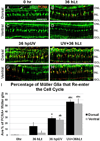
Figure 3. Müller glial cell proliferation in various light treatments
EGFP-positive Müller glial cells in the dorsal (A, B, E, F) and ventral (C, D, G, H) halves of the retina from adult Tg(gfap:egfp)/alb zebrafish (Green) co-labeled with PCNA (Red). A. Untreated (0 hr) dorsal retina, showing EGFP-positive Müller glial cells with somas located in the INL and a processes extending to beyond the ONL and GCL. Müller glial cells in 0 hr retinas are not PCNA-positive. B. A subset of Müller glial cells are PCNA-positive in the dorsal retina following 36 hours of constant bright light treatment (36 hLt). C. 0 hr ventral retina, showing PCNA-negative Müller glial cells. D. 36 hLt dorsal retina, showing PCNA-negative Müller glial cells. E. 48 hours after a 30 minute UV exposure (36 hpUV), a subset of Müller glial cells are PCNA-positive in the dorsal retina. F. After a 30 minute exposure to UV light followed by 48 hours of constant bright light (UV+48 hLt), the majority of Müller glial cells are PCNA-positive in the dorsal retina. G. 36 hpUV retina, showing PCNA-positive Müller glial cells in the dorsal half of the ventral retina. H. UV+48 hLt retina, showing the majority of Müller glial cells are PCNA-positive in the ventral retina. I. Quantification of the percentage of PCNA-positive Müller glial cells in the entire dorsal and ventral halves of the retina in untreated and light-treated retinas. Significant differences between groups (p < 0.05) is indicated as: “a” for compared with Untreated (0 hr); “b” for compared with 36 hLt; and “c” for compared with 36 hpUV. Scale bar in panel A represents 50 microns.
Figure 4
Figure 4. PCNA immunolocalization following various light damage paradigms
A. Untreated (0 hr) dorsal retina, showing the absence of PCNA immunolocalization in the inner nuclear layer (INL). B – D. PCNA immunolocalization in the INL and ONL of the dorsal retina, following each of the light damage paradigms (48 hLt, 48 hpUV, and UV+48 hLt). E. 0 hr ventral retina, showing PCNA immunolocalization in the ONL, but not in the INL. F – G. Following 48 hLt (F) and 48 hpUV (G), isolated PCNA-positive cells are observed in the ONL and INL of the ventral half of the retina, however, the proliferation response if markedly less than that observed in the corresponding dorsal half of the retina. H. Ventral retinas from UV+48 hLt possess large numbers of PCNA-positive cells in the INL and ONL. Scale bar in panel A represents 50 microns.
Figure 5
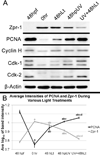
Figure 5. Western blot analysis following various light damage paradigms
A. Expression levels of Zpr-1, PCNA, Cyclin H, Cdk-1, Cdk-2, and β-actin were determined in 48 hour post-fertilization embryos (48 hpf), untreated adult retinas (0 hr), and following the various light damage paradigms (48 hLt, 48 hpUV, and UV+48 hLt). Zpr-1 was not detected at 48 hpf since double cones are not present at this early stage in development. Zpr-1 expression decreased from 0 hr→48 hLt→48 hpUV→UV+48 hLt, representing a progressive loss of double cones. Conversely, PCNA and Cyclin H levels increased from 0 hr→48 hLt→48 hpUV→UV+48 hLt. Two bands were observed for Cdk-1 and Cdk-2, most likely representing phosphorylated and unphosphorylated forms of each protein. For Cdk-1, the upper band was not detected at 0 hr, and both the upper and lower bands increased from 0 hr→48 hLt→48 hpUV→UV+48 hLt. For Cdk-2, only the upper band was detected at 0 hr (and slightly at 48 hLt), and the lower band increased from 0 hr→48 hLt→48 hpUV→UV+48 hLt. B. Graphic representation of the average log10 band density of Zpr-1 and PCNA expression. Significant differences between groups (p < 0.05) is indicated as: “a” for compared with 48 hpf; “b” for compared with untreated (0 hr); “c” for compared with 48 hLt’ and “d” for compared with 48 hpUV.
Figure 6
Figure 6. Anterior/Posterior differences in light-induced proliferation response
Cryosections of the dorsal half of the retina, taken from the anterior (A–D), central (E–L), or posterior regions (M–P) of the retina were immunolabeld with PCNA (red). In untreated (0 hr) retinas (A, E, I, M), PCNA-positive cells were occasionally observed as rod precursors in the outer nuclear layer (ONL). Following 48 hours of constant bright light (48 hLt; panels B, F, J, and N), PCNA-positive cells were observed in the inner nuclear layer (INL) and ONL in sections from the anterior and central retina, but not the posterior retina. In addition, the proliferation response was much more robust in the anterior-most section (B). 48 hours after a 30 minute UV exposure (48 hpUV; panels C, G, K, and O), clusters of PCNA-positive cells were observed in the INL and ONL in sections from the anterior and central retina. The posterior retina (O), in contrast, possessed isolated PCNA-positive cells in both the INL and ONL, indicative of a more subdued proliferative response. After a 30 minute exposure to UV light followed by 48 hours of constant bright light (UV+48 hLt; panels D, H, L, and P), large clusters of PCNA-positive nuclei were present throughout the anterior, central, and posterior retina. Scale bar in panel A is 50 microns.
Figure 7
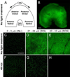
Figure 7. Orientation and imaging of flatmounted retinas
A. Schematic representation of the orientation used for these studies on flatmounted retinas. Anterior is to the left and posterior is to the right. B. Stereofluorescent image of an untreated, flatmounted Tg(nrd:egfp)/alb retina, oriented as shown in panel A. C – H. Confocal images taken at various focal planes in the anterior retina (equidistant between the dorsal and ventral halves) revealed the inner retina (INL), rod inner segments (RIS), and rod outer segments (ROS). C. EGFP-positive bipolar cells were detected in the INL of untreated retinas. D. EGFP-positive RIS exhibited the characteristic honeycomb pattern in untreated retinas. E. EGFP-positive ROS were undamaged in untreated retinas. F. Following 48 hours of constant bright light (48 hLt), EGFP-positive bipolar cells were present in the INL and appeared hypertrophied. G. EGFP-positive RIS in 48 hLt retinas were damaged (note large EGFP-negative areas) and disorganized. H. ROS were not detected in 48 hLt retinas. Scale bar in panel B represents 450 microns. The scale bar in panel C (C–H) represents 100 microns.
Figure 8
Figure 8. Confocal microscopy of rod inner segments in flatmounted retinas reveal anterior/posterior differences in response to various light-damage paradigms
Confocal images of EGFP-positive rod inner segments (RIS) were obtained from the central anterior (A–D) and central posterior (E–H) portion of flatmounted Tg(nrd:egfp)/alb retinas. Untreated (0 hr) retinas (A, E) showed the characteristic honeycomb pattern of RIS (note the inset in panel A) in both the anterior and posterior halves of the retina. Following 48 hours of constant bright light (48 hLt; panels B and F), the RIS in the anterior half of the retina were severely disrupted (B), but remained largely intact in the posterior half of the retina (F). 48 hours after a 30 minute UV exposure (48 hpUV; panels C and G), RIS in both the anterior and posterior halves of the retina were severely damaged. After a 30 minute exposure to UV light followed by 48 hours of constant bright light (UV+48 hLt; panels D and H), RIS in both the anterior and posterior halves of the retina were severely damaged. Compared with the other treatments, the anterior half of the UV+48 hLt retinas appeared to sustain the greatest damage to RIS (compare panel D with all other panels). I. Quantification of the fluorescent intensity of EGFP-positive RIS relative to RIS in the posterior portion of the retina. Significant differences between groups (p < 0.05) is indicated as: “a” for significantly less than the untreated retina; “b” for significantly less than the posterior half of the corresponding treatment group; and “c” for significantly less than all other points. Scale bar in panel A represents 100 microns.

References

    1. Bernardos RL, Barthel LK, Meyers JR, Raymond PA. Late-stage neuronal progenitors in the retina are radial Muller glia that function as retinal stem cells. J Neurosci. 2007;27:7028–7040. - PMC - PubMed
    1. Chang GQ, Hao Y, Wong F. Apoptosis: final common pathway of photoreceptor death in rd, rds, and rhodopsin mutant mice. Neuron. 1993;11:595–605. - PubMed
    1. Cicerone CM. Cones survive rods in the light-damaged eye of the albino rat. Science. 1976;194:1183–1185. - PubMed
    1. Devault A, Fesquet D, Cavadore JC, Garrigues AM, Labbe JC, Lorca T, Picard A, Philippe M, Doree M. Cyclin A potentiates maturation-promoting factor activation in the early Xenopus embryo via inhibition of the tyrosine kinase that phosphorylates cdc2. J Cell Biol. 1992;118:1109–1120. - PMC - PubMed
    1. Faktorovich EG, Steinberg RH, Yasumura D, Matthes MT, LaVail MM. Photoreceptor degeneration in inherited retinal dystrophy delayed by basic fibroblast growth factor. Nature. 1990;347:83–86. - PubMed

Publication types

MeSH terms

LinkOut - more resources