Comparative Study
. 2012 Sep;26(9):2114-23.
doi: 10.1038/leu.2012.71. Epub 2012 Mar 19.

Constitutive activation of p38 MAPK in tumor cells contributes to osteolytic bone lesions in multiple myeloma

Affiliations
Comparative Study

Constitutive activation of p38 MAPK in tumor cells contributes to osteolytic bone lesions in multiple myeloma

J Yang et al. Leukemia. 2012 Sep.

Erratum in

  • Leukemia. 2015 Feb;29(2):515

Abstract

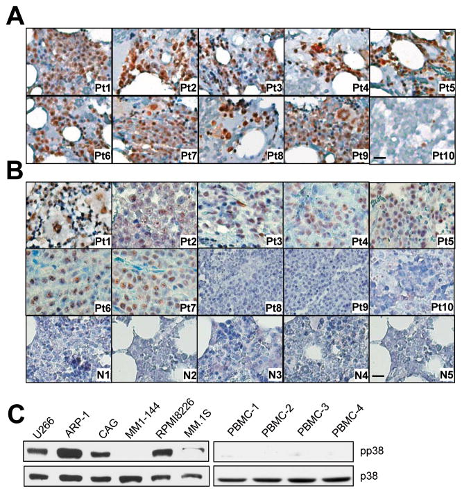
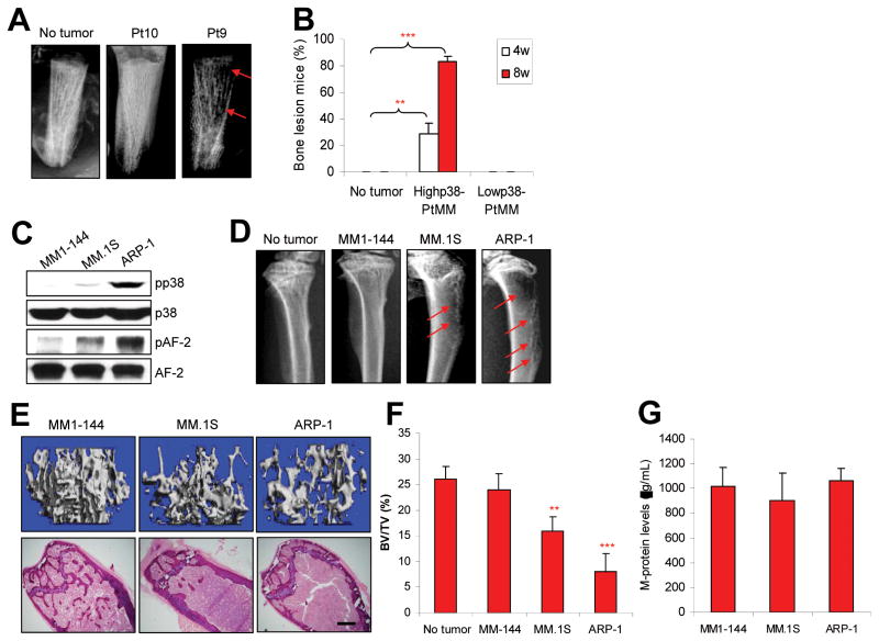
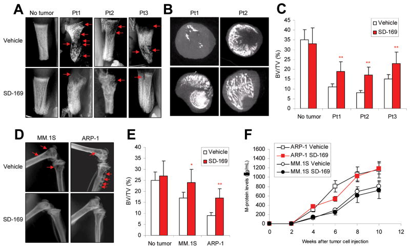
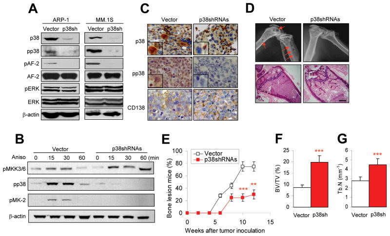
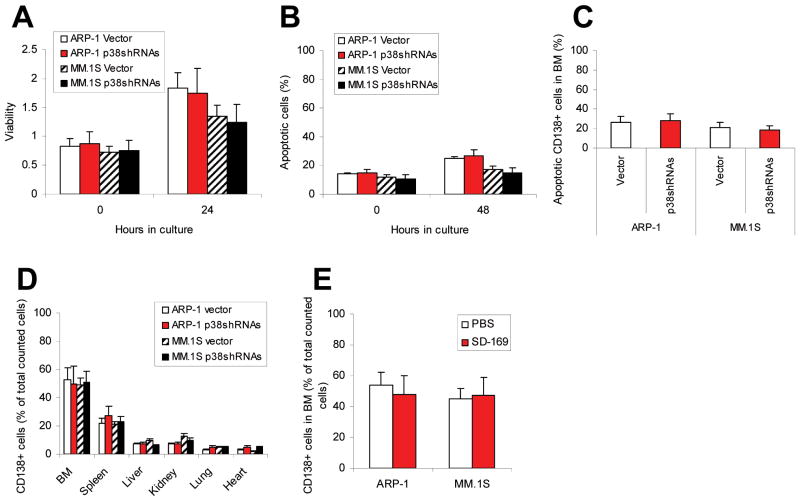
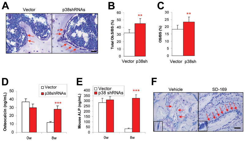
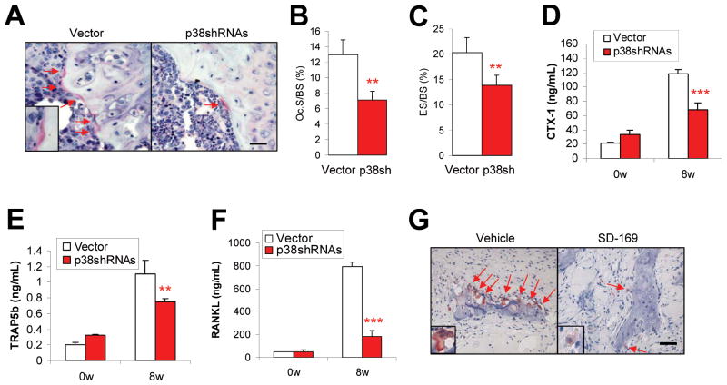
Bone destruction is a hallmark of multiple myeloma and affects more than 80% of patients. However, current therapy is unable to completely cure and/or prevent bone lesions. Although it is accepted that myeloma cells mediate bone destruction by inhibition of osteoblasts and activation of osteoclasts, the underlying mechanism is still poorly understood. This study demonstrates that constitutive activation of p38 mitogen-activated protein kinase in myeloma cells is responsible for myeloma-induced osteolysis. Our results show that p38 is constitutively activated in most myeloma cell lines and primary myeloma cells from patients. Myeloma cells with high/detectable p38 activity, but not those with low/undetectable p38 activity, injected into severe combined immunodeficient (SCID) or SCID-hu mice caused bone destruction. Inhibition or knockdown of p38 in human myeloma reduced or prevented myeloma-induced osteolytic bone lesions without affecting tumor growth, survival, or homing to bone. Mechanistic studies showed that myeloma cell p38 activity inhibited osteoblastogenesis and bone formation and activated osteoclastogenesis and bone resorption in myeloma-bearing SCID mice. This study elucidates a novel molecular mechanism-activation of p38 signaling in myeloma cells-by which myeloma cells induce osteolytic bone lesions, and indicates that targeting myeloma cell p38 may be a viable approach to treating or preventing myeloma bone disease.


Conflict of interest statement

Disclosure of Potential Conflicts of Interest

No potential conflicts of interest were disclosed.

Figures

Figure 1
Figure 1
Constitutive activation of p38 in myeloma cells. Representative images of immunohistochemical staining for pp38 in (A) bone marrow biopsy specimens of 10 randomly selected patients with newly diagnosed myeloma (Pt1–Pt10) and (B) a tissue array containing bone marrow samples of 10 myeloma patients (Pt1–Pt10) and 11 healthy donors. (Only five samples, N1–N5; are shown. The other six samples also stained negatively for pp38.) Scale bar, 10 μM. Western blot analysis showing the levels of phosphorylated p38 (pp38) and nonphosphorylated p38 (p38) in (C) six myeloma cell lines and in (D) three normal PBMCs. Representative results of three independent experiments are shown.
Figure 2
Figure 2
Myeloma cells with high/detectable p38 activity cause bone lesions in mouse models. (A) Representative radiographic images show lytic bone lesions in the implanted human bones of SCID-hu mice bearing primary myeloma cells from one (patient 10; pt10) of four patients with low or undetectable p38 or one (patient 9; pt9) of eight patients with high/detectable p38 activity. (B) Percentages of mice with bone lesions, detected by radiographs, at 4 and 8 weeks after injection ofprimary myeloma cells with high/detectable p38 activity from eight patients or with undetectable pp38 activity from four patients. (C) Western blot analysis shows the levels of phosphorylated p38 (pp38) and nonphosphorylated p38 (p38) and AF-2 in MM1-144, MM.1S, and ARP-1 myeloma cells. (D) Representative radiographic images show lytic bone lesions in the distal femurs of SCID mice (10 per each cell line) injected with ARP-1, MM.1S, or MM1-144 myeloma cells. Tumor-free mice (no tumor) served as controls. Red arrows indicate osteolytic lesions. (E) Representative μ-CT images (upper panels) and histologic examinations (lower panels) and (F) quantitative analyses show the trabecular bone volume density (BV/TV) in distal femurs of SCID mice bearing ARP-1, MM.1S, or MM1-144 myeloma. ** P < 0.01 to *** P < 0.001, compared with tumor-free mice. Scale bars: 1 mm. (G) ELISA showing no difference in the levels of M-protein in serum of mice injected with ARP-1, MM.1S, or MM1-144 myeloma cells at week 8 after tumor injection. Similar results were obtained at week 4 and week 6 after tumor injection.
Figure 3
Figure 3
Administration of p38 inhibitor reduces myeloma-induced osteolytic bone lesions. In vivo injection of p38 specific inhibitor SD-169 significantly reduced myeloma-induced bone lesions in fetal human bones implanted into SCID-hu mice bearing a primary myeloma xenograft; lesions were detected by (A) radiography and (B) μ-CT scanning. (C) SD-169 also reduced bone lesions in distal femurs of ARP-1- or MM.1S-bearing SCID mice. (D) Tumor burden was measured as circulating human Ig in SCID mice inoculated with ARP-1 or MM.1S cells treated without or with p38 inhibitor SD-169. CD138+ primary myeloma cells were isolated from three myeloma patients (Pt1, Pt2, and Pt3) with high/detectable p38 activity and injected into the implanted human bones of SCID-hu mice. Myeloma ARP-1 or MM.1S cells were injected intravenously into SCID mice (10 per each cell line). When circulating M-protein levels reached 10 μg/ml, mice were fed daily with SD-169 (10 mg/kg) or an equal volume of PBS (vehicle controls) for a total of 30 days. Arrows indicate osteolytic bone lesions.
Figure 4
Figure 4
Knockdown of tumor cell p38 activity by shRNAs reduces myeloma-induced osteolytic bone lesions. Myeloma cells were stably transfected with retrovirus containing control vector or p38-shRNAs to knock down p38 activity. Western blot analysis shows (A) levels of phosphorylated p38 (pp38), nonphosphorylated p38 (p38), nonphosphorylated or phosphorylated AF-2 (a p38 downstream kinase), and nonphosphorylated or phosphorylated ERK1/2 in vector-(vector) or p38 shRNA-ARP-1 or -MM.1S cells, and (B) levels of phosphorylated MKK3/6 (pMKK3/6, upstream kinase of p38), pp38, and phosphorylated MAPKPK-2 (pMK-2, downstream kinase of p38) in vector- or p38 shRNA-ARP-1 (p38shRNAs) or -MM.1S (data not shown) cells treated with 10 nM of anisomycin (Aniso) at different time points (0, 15, 30, 60 minutes). β-actin protein levels served as loading controls. (C) Immunohistochemical staining shows the expression of p38, pp38, and CD138 (myeloma cell marker) in bone sections of SCID mice injected with vector- (vector) or p38 shRNA-ARP-1 cells. The vector-ARP-1 or p38 shRNA-ARP-1 cells were intravenously injected into SCID mice. Sections of distal femur taken from mice killed 4 weeks after tumor inoculation were immunostained with antibodies against p38, pp38, and CD138. Scale bar, 10 μM. (D) Representative radiographic images (upper panels) show osteolytic lesions in the distal femurs and histologic examinations (lower panels) show the densities of trabecular bones in the bone marrow of SCID mice (10 per group) injected with vector- or p38 shRNA-ARP-1 cells and killed 8 weeks after tumor injection. Arrows indicate osteolytic bone lesions. (E) Percentages of mice with bone lesions, detected by radiographs, are shown at different time intervals after injection with vector- or p38shRNA-ARP-1 cells. Analysis of bone sections by μ-CT scanning shows (F) greater trabecular bone volumes and (G) larger densities inthe distal femurs of SCID mice injected with p38 shRNA-ARP-1 cells than in those injected with vector-ARP-1 cells. **P ≤ 0.01; ***P ≤ 0.001.
Figure 5
Figure 5
Disruption of myeloma cell p38 activity has no effect on tumor growth or survival or on the ability of myeloma cells to home to bones. Shown are (A) viability of vector-or p38 shRNA-ARP-1 or -MM.1S cells (p38shRNAs) examined byMTT assayat 0 and 24 hour culture; and (B) percentages of apoptotic vector- or p38 shRNA-ARP-1 or -MM.1S cells examined by Annexin-V binding assay at 0 and 48 hour cultures. Results represent average values from five independent experiments. (C) In situ TUNEL assay results show the percentages of apoptotic CD138+ tumor cells in the bone marrow of SCID mice injected with vector- (vector) or p38 shRNA-ARP-1 or -MM.1S cells. Flow cytometry results show tissue-infiltrating CD138+ human myeloma cells in (D) different organs, including bone marrow (BM), spleen, liver, kidney, heart and lung, of SCID mice injected with vector- (vector) or p38 shRNA-ARP-1 or -MM.1S cells or (E) bone marrow of SCID mice injected with parental ARP-1 or MM.1S cells and treated with p38 inhibitor SD-169 or PBS. In the experiments, myeloma cells were injected intravenously into SCID mice (10 per each cell line). When circulating M-protein levels reached 10 μg/ml, mice were fed daily with SD-169 (10 mg/kg) or an equal volume of PBS (vehicle controls) for a total of 30 days. After treatment, bone marrow cells were flushed out and CD138+ human myeloma cells were identified with flow cytometry. The results represent average values from three independent experiments of five mice per group.
Figure 6
Figure 6
Activated tumor cell p38 inhibits osteoblastogenesis in vivo. Shown are (A) representative images of toluidine blue staining showing the numbers of osteoblasts, and the results of quantitative analysis of osteoblast parameters, including (B) total osteoblast numbers on the surface and (C) osteoid percentage of trabecular bone of SCID mice bearing vector- or p38 shRNA-ARP-1 myeloma. ELISA showed levels of circulating (D) osteocalcin or (E) ALP in mice prior to (week 0; 0w) and 8 weeks (8w) after injection with controls cells or p38 shRNA-ARP-1 cells. (F) Immunohistochemical staining shows the numbers of osteoblasts present at week 8 on the surface of human trabecular bone implanted into primary myeloma-xenografted SCID-hu mice treated without (vehicle) or with p38 inhibitor SD-169. Arrows indicate osteoblasts. Generation of osteoblasts from normal mesenchymal stem cells in osteoblast medium with addition of conditioning medium of vector or p38 shRNA-ARP-1 cells (p38SH). Representative results from four independent experiments are shown. **P ≤ 0.01; ***P ≤ 0.001.
Figure 7
Figure 7
Activated tumor cell p38 enhances osteoclastogenesis in vivo. Shown are (A) representative images and numbers of TRAP-positive osteoclasts on murine trabecular bone (scale bars in larger windows: 50 μm; in smaller windows: 10 μm), and quantitative analysis of osteoclast parameters such as (B) percentages of osteoclast-mediated erosion of bone surface (ES/BS) and (C) numbers of osteoclasts present on bone surface (Oc. S/BS) in bone sections of SCID mice bearing vector- or p38 shRNA-ARP-1 (p38shRNAs/p38SH) myeloma at week 8 after tumor inoculation. Shown are levels of circulating (D) CTX-1, (E) TRAP5b, and (F) RANKL in mice injected with vector- or p38 shRNA-ARP-1 cells at 0 week (0w) or 8 weeks (8w) after tumor inoculation. (G) Immunohistochemical staining shows the numbers of TRAP-positive osteoclasts present on human trabecular bone surfaces of primary myeloma-bearing SCID-hu mice treated without (vehicle) or with p38 inhibitor SD-169 at week 8 after tumor injection. Arrows indicate osteoclasts. Representative results from four independent experiments are shown. **P ≤ 0.01; ***P ≤ 0.001.

Similar articles

Cited by

References

    1. Roodman GD. Myeloma bone disease: pathogenesis and treatment. Oncology (Williston Park) 2005 Jul;19(8):983–984. 986. - PubMed
    1. Tian E, Zhan F, Walker R, Rasmussen E, Ma Y, Barlogie B, et al. The role of the Wnt-signaling antagonist DKK1 in the development of osteolytic lesions in multiple myeloma. N Engl J Med. 2003 Dec 25;349(26):2483–2494. - PubMed
    1. Giuliani N, Rizzoli V, Roodman GD. Multiple myeloma bone disease: Pathophysiology of osteoblast inhibition. Blood. 2006 Dec 15;108(13):3992–3996. - PubMed
    1. Wang S, Hong S, Yang J, Qian J, Zhang X, Shpall E, et al. Optimizing immunotherapy in multiple myeloma: Restoring the function of patients’ monocyte-derived dendritic cells by inhibiting p38 or activating MEK/ERK MAPK and neutralizing interleukin-6 in progenitor cells. Blood. 2006 Dec 15;108(13):4071–4077. - PMC - PubMed
    1. Wang S, Yang J, Qian J, Wezeman M, Kwak LW, Yi Q. Tumor evasion of the immune system: inhibiting p38 MAPK signaling restores the function of dendritic cells in multiple myeloma. Blood. 2006 Mar 15;107(6):2432–2439. - PMC - PubMed

Publication types

MeSH terms

Substances