. 2012 Mar 18;14(4):355-65.
doi: 10.1038/ncb2466.

Telomeric DNA damage is irreparable and causes persistent DNA-damage-response activation

Affiliations

Telomeric DNA damage is irreparable and causes persistent DNA-damage-response activation

Marzia Fumagalli et al. Nat Cell Biol. .

Erratum in

  • Nat Cell Biol. 2012 May;14(5):555

Abstract

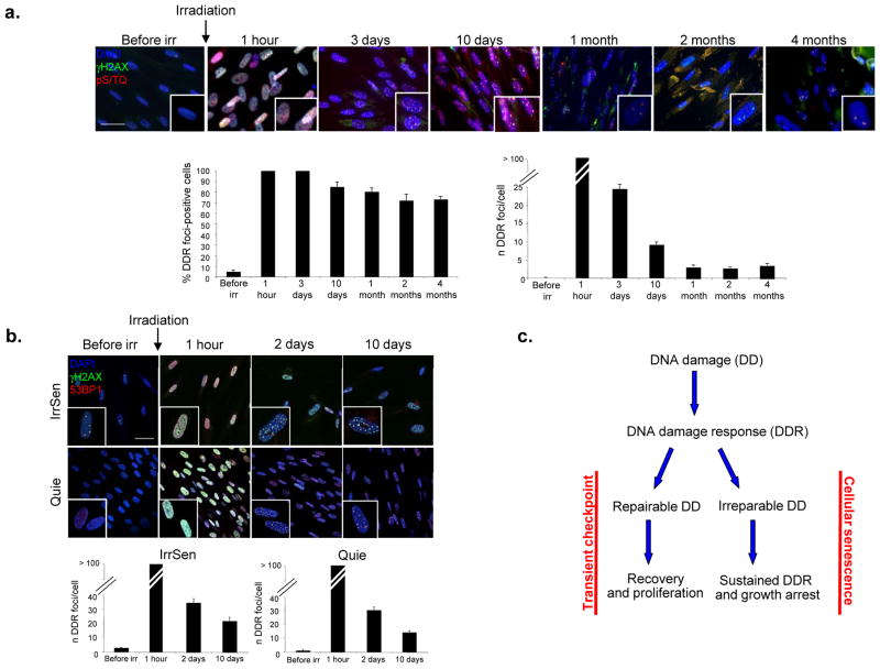
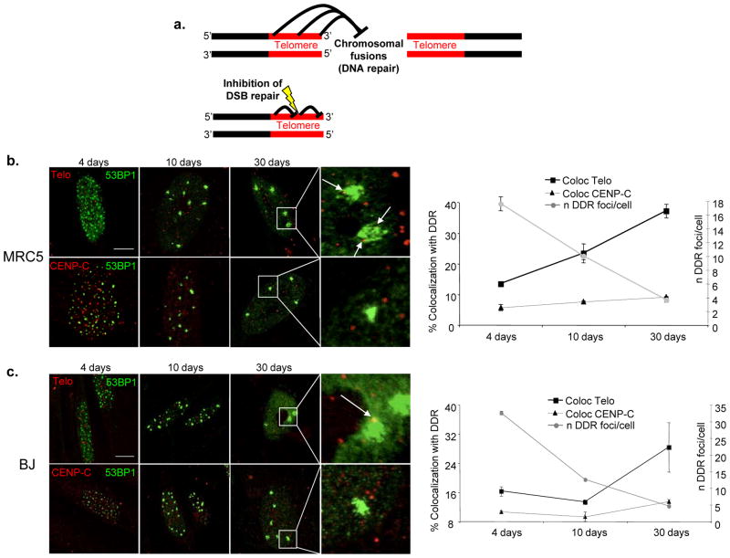
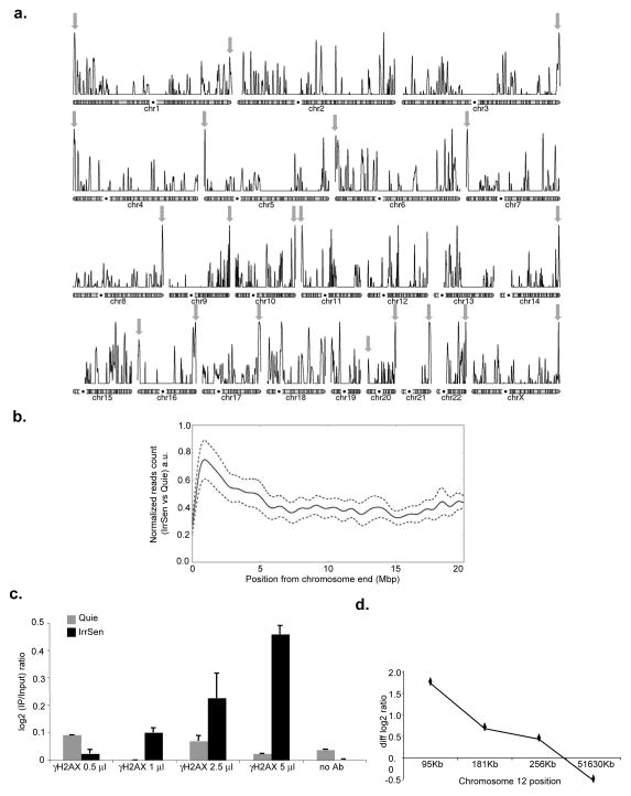
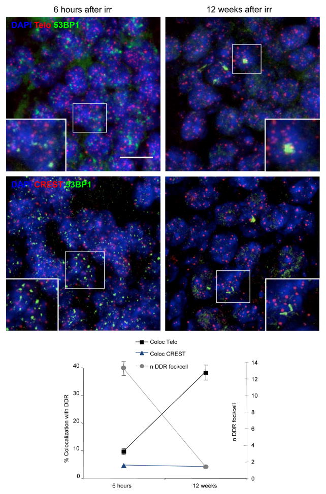
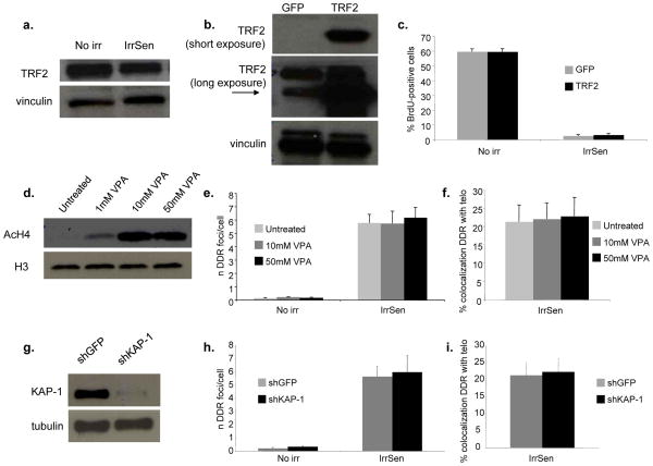
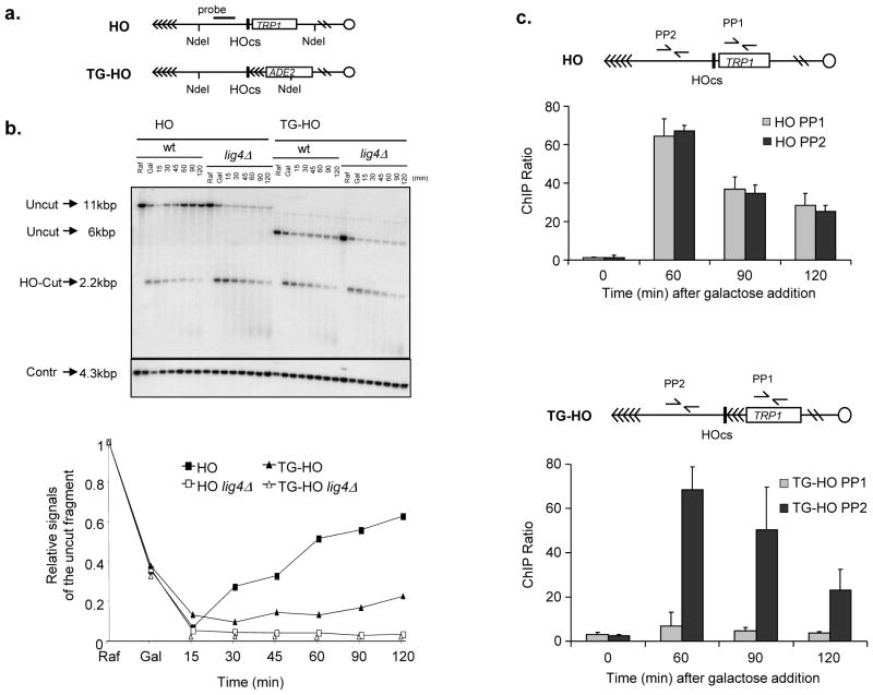
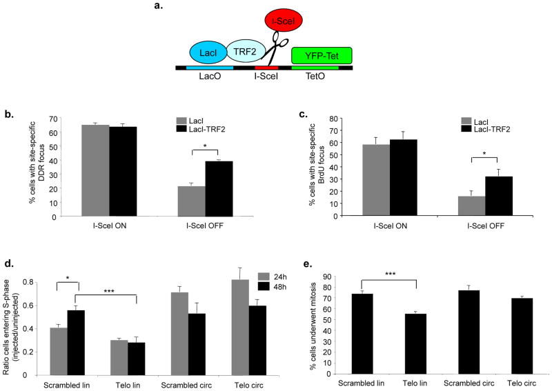
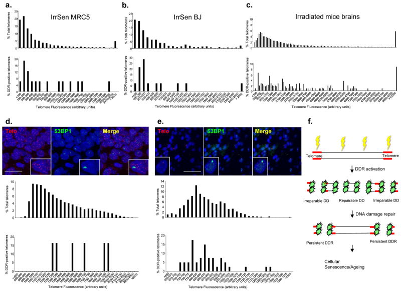
The DNA-damage response (DDR) arrests cell-cycle progression until damage is removed. DNA-damage-induced cellular senescence is associated with persistent DDR. The molecular bases that distinguish transient from persistent DDR are unknown. Here we show that a large fraction of exogenously induced persistent DDR markers is associated with telomeric DNA in cultured cells and mammalian tissues. In yeast, a chromosomal DNA double-strand break next to a telomeric sequence resists repair and impairs DNA ligase 4 recruitment. In mammalian cells, ectopic localization of telomeric factor TRF2 next to a double-strand break induces persistent DNA damage and DDR. Linear, but not circular, telomeric DNA or scrambled DNA induces a prolonged checkpoint in normal cells. In terminally differentiated tissues of old primates, DDR markers accumulate at telomeres that are not critically short. We propose that linear genomes are not uniformly reparable and that telomeric DNA tracts, if damaged, are irreparable and trigger persistent DDR and cellular senescence.


Conflict of interest statement

The authors declare no conflict of interests.

Figures

Figure 1
Figure 1. IR induces persistent DDR activation and cellular senescence
a. IR generates persistent DDR. Top, images show DDR foci induction and resolution in early passage quiescent (contact-inhibited) BJ human fibroblasts following exposure to 20 Gy IR. Persistent DDR, in the form of γH2AX and pS/TQ foci, is still detectable even 4 months after IR. Bottom, bar graphs show the fraction of γH2AX foci-positive cells (± s.e.m.) (on the left) and the average number of γH2AX foci (± s.e.m.) per cell (on the right), at the indicated time points. More than 100 discrete foci cannot be counted accurately due to their proximity (1 hour time point). (For the quantification shown, around 100 cells per time point were analysed; scale bar = 20 μm) b. IrrSen cells are able to resolve additional IR-induced (10 Gy) DNA damage to an extent similar to Quie cells, as shown by the comparable kinetics of resolution of 53BP1 and γH2AX foci per cell over time after IR. (For the quantification shown, around 100 cells per time point were analysed; scale bar = 50 μm) c. Model, two opposite outcomes are possible upon DNA damage generation.
Figure 2
Figure 2. Persistent DDR is preferentially associated with telomeric DNA
a. A model of inhibition of DNA repair at telomeres. Top, Telomeric repeats prevent DNA end joining (DNA repair by NHEJ) at their distal end in order to prevent chromosomal fusions. Bottom, in the same manner, stretches of telomeric repeats may prevent DNA end joining (DNA repair by NHEJ) of DNA damage generated within repeats across the telomere length. b–c. Persistent DDR foci colocalize with telomeres in IrrSen human diploid fibroblasts (HDFs). Left, representative pictures of colocalizations between DDR, detected as 53BP1 foci, and telomeres, detected using a telomeric PNA probe (Telo), or centromeres, detected by antibodies raised against a centromeric protein (CENP-C), at the indicated time points following IR in MRC5 (b.) (scale bar = 10 μm) and in BJ (c.) (scale bar = 20 μm) cells. 30 days after IR, IrrSen HDFs show the highest degree of colocalizations between 53BP1 and the telomeric PNA probe. Arrows indicate telomeric signals colocalizing with 53BP1 foci. Right, graphs show the percentage of colocalizations (± s.e.m.) between 53BP1 foci and telomeric (Telo) or centromeric (CENP-C) regions, and the average number of 53BP1 foci per cell (± s.e.m.) at the indicated time points after IR. (For the quantifications shown, around 50–200 cells per time point were analysed).
Figure 3
Figure 3. Persistent DDR is physically associated with telomeric DNA
a. Chromosomal view of the enrichment of γH2AX in IrrSen vs Quie cells. Individual chromosomes representation of the enrichment of γH2AX in IrrSen BJ hTERT cells. Grey arrows indicate the highest peak of each chromosome arm within 5 Mbp from a chromosome end. b. Enrichment of γH2AX in IrrSen vs Quie cells at chromosome ends. Solid line represents the accumulation pattern of γH2AX over 20 Mbp from the chromosomes end. The profile shows an enrichment peak at the most terminal region (0–5 Mbp). The enrichment is significantly different when compared to the accumulation in a more internal region (15–20 Mbp), (Mann-Whitney-U p-value = 0, n=2.3e6). 95% credibility intervals are shown as dashed lines; a.u. means arbitrary units. c. Increasing amounts of antibodies against γH2AX immunoprecipitate increasing amounts of subtelomeric DNA from IrrSen BJ cells compared to non-irradiated Quie cells, as assayed on telomere 12p by ChIP and qPCR using a primer pair distant 95 Kb from chromosome end. Triplicate qPCR reactions were carried out and the averaged results are plotted as the log2 ratio between IP and input (n = 2). d. γH2AX and subtelomeric DNA are physically associated in IrrSen BJ cells. Enrichment of γH2AX was assayed on telomere 12p by ChIP and qPCR with previously independently-validated primer pairs at indicated increasing distances from the chromosome end. Graph shows subtracted log2 ratios between IP and input of IrrSen minus Quie cells (diff log2 ratio). Triplicate qPCR reactions were carried out and the averaged results are plotted (n = 2).
Figure 4
Figure 4. IR generates persistent DDR at telomeres in vivo
DDR, in the form of 53BP1 foci, telomeres, as detected by a telomeric PNA probe (Telo), or centromeres, visualized with antibodies against centromeric proteins (CREST), were analysed in hippocampal neurons of irradiated adult mice at the indicated time points post IR. Top, representative pictures show activation of DDR immediately after IR, persistence of individual foci and their colocalizations with telomeres at 12 weeks (last time point analyzed) after IR. Bottom, graph shows the average number of 53BP1 foci per cell in DDR-positive cells and the percentage of colocalizations (± s.e.m.) between 53BP1 foci and telomeric DNA (Telo) or centromeres (CREST). (For the quantification shown, around 400 cells per time point were analysed: n = 3: scale bar = 10 μm).
Figure 5
Figure 5. TRF2 overexpression does not prevent senescence establishment and heterochromatin disruption does not prevent the persistence of DDR at telomeres
a. TRF2 expression is not altered in IrrSen cells. Immunoblot shows TRF2 protein levels in IrrSen BJ hTERT, compared to non-irradiated cells (No irr). Vinculin was used as a loading control. b. Immunoblot showing TRF2 expression in BJ hTERT cells infected with either TRF2- or GFP-expressing lentiviruses. Vinculin was used as a loading control. c. TRF2 overexpression does not prevent senescence establishment. TRF2 and GFP overexpressing BJ hTERT cells were irradiated (20 Gy) and analyzed 30 days later. Bar graphs show the percentage of BrdU-positive cells (± s.e.m.) (For the quantification shown, around 400 cells per sample were analysed). d. Heterochromatin disruption by VPA treatment does not significantly affect the number of persistent DDR foci and their colocalization with telomeres. Immunoblot shows the increased levels of acetylated histone H4 (AcH4) in BJ hTERT cells treated with the indicated concentration of VPA, compared to untreated control. H3 was used as a loading control. e. VPA-treated cells were irradiated with 20 Gy and analyzed 30 days later. Bar graphs show the number of 53BP1 foci per cell and (f.) the percentage of 53BP1 foci colocalizing with a telomeric PNA probe (± s.e.m.), in cells treated with the indicated doses compared to untreated control. (For the quantification shown, around 30–100 cells per sample were analyzed). g. KAP-1 knock down does not significantly affect the number of persistent DDR foci and their colocalization with telomeres. Immunoblot shows the expression of KAP-1 in shKAP-1 and shGFP BJ hTERT. Tubulin was used as a loading control. h. Cells were irradiated with 20 Gy and analyzed 30 days later. Bar graphs show the number of 53BP1 foci per cell and (i.) the percentage of 53BP1 foci colocalizing with a telomeric PNA probe (± s.e.m.). (For the quantification shown, around 30–100 cells per sample were analyzed).
Figure 6
Figure 6. Lack of repair of a chromosomal DSB adjacent to telomeric DNA repeats and impaired ligase 4 recruitment
a. Schematic of the HO (RMY169 and RMY169 lig4Δ ) or the TG-HO (UCC5913 and UCC5913 lig4Δ ) system on yeast chromosome VII. HOcs represents the cutting site for the HO endonuclease, which is flanked by 81 bp TG sequence (represented by arrows) in the TG-HO strain. b. HO-cut formation and repair in strains carrying the HO or the TG-HO system at chromosome VII. G1-arrested cell cultures in YEP+raffinose (Raf) were supplemented with galactose (Gal) to induce HO expression. After 1 hour of induction, cells were washed and transferred to YEPD in the presence of α-factor to maintain the G1 arrest. Genomic DNA prepared at different time points after galactose removal were subjected to southern blot analysis with the probe indicated in (a). Top, the probe reveals an uncut fragment in the absence of HO-cut or after the break had been repaired by NHEJ (Uncut), whereas the HO-induced DSB results in the formation of an HO-cut DNA fragment (HO-Cut). (Contr) represents the loading control. Bottom, quantification of a representative experiment. Three independent experiments were performed with similar results. c. Ligase 4 is efficiently recruited to the DSB site only when it is not flanked by telomeric repeats. HO expression was induced at time zero by galactose addition to G1-arrested cells carrying the HO (top) or TG-HO (bottom) system. Lig4 recruitment was analyzed by ChIP and qPCR. Data are expressed as relative fold enrichment (± s.d.) of PP1 or PP2 over CON signal after normalization to input signals for each primer set (n = 4).
Figure 7
Figure 7. Ectopic TRF2 modulates DNA repair and DDR focus persistence, and exposed telomeric DNA ends cause a prolonged checkpoint
a. Schematic of the integrated locus studied in NIH 2/4 cells. Upon transfection, LacI or LacI-TRF2 binds to the lactose operator (LacO) repeats, YFP-Tet binds to the tetracycline operator (TetO) repeats, and RFP-I-SceI-GR cuts the specific site between the two sets of repeats. b. Quantification of cells positive for γH2AX at the I-SceI-locus (± s.e.m.) expressing LacI or LacI-TRF2, as detected by immunofluorescence confocal microscopy. I-SceI ON corresponds to 3 hours after RFP-I-SceI-GR induction, I-SceI OFF corresponds to 24 additional hours after removal of inducing agent. I-SceI site was detected as a distinct focus double-positive for YFP-Tet and anti-LacI antibody signals (* p value < 0.05; for the quantification shown, around 100 cells per sample were analyzed; n = 2). c. Quantification of cells positive for a BrdU signal (± s.e.m.) at the I-SceI-locus expressing LacI or LacI-TRF2, as detected by BrdU immunostaining under non-denaturing conditions and confocal microscopy. Values were normalized on the fraction of cells that had incorporated BrdU. (* p value < 0.05; for the quantification shown, around 100 cells per sample were analyzed; n = 2). d. Linearized telomeric DNA triggers a prolonged cell cycle arrest. Bar graph shows the ratio (± s.e.m.) between the percentages of injected cells that underwent DNA synthesis (assayed by BrdU incorporation) and the percentages of uninjected cells in the same experiment, at 24 or 48 hours after microinjection (* p-value < 0.05; *** p-value < 0.001; for the quantification shown, around 200–400 cells per time point, per DNA type were analyzed; n = 3). e. Linearized telomeric DNA impedes cell proliferation. Bar graph shows the percentages (± s.e.m.) of cells that underwent mitosis at 48 hours post-microinjection (*** p-value < 0.001; for the quantification shown, around 200 cells per DNA type were analyzed; n = 3). Passage through mitosis was monitored by detection in the cytoplasm of nucleus-injected IgG 48 hours after microinjection, as detected by immunofluorescence.
Figure 8
Figure 8. Persistent DDR accumulates at telomeres independently of their lengths, also in ageing primates
a.–c. Association of persistent DDR at the telomeres is not triggered by telomere shortening. Relative distribution of total telomeres lengths (upper histograms) and of 53BP1 focus-positive telomeres lengths (lower histograms) according to telomeric probe signal intensity (Telomere Fluorescence Arbitrary Units), in IrrSen HDFs (MRC5, a., and BJ, b.; approximately 1000 telomeres per sample were analyzed) and (c.) in in vivo mouse hippocampal neurons 12 weeks after IR (telomeres of approximately 200 cells per sample were analyzed; n = 3). DDR in hippocampal neurons (d.) and in liver hepatocytes (e.) of ageing baboons is not preferentially associated with the shortest telomeres. Top, representative images of telomeric PNA probe (Telo) and 53BP1 foci in hippocampal neurons (scale bar = 20 μm) and liver hepatocytes (scale bar = 50 μm) of aged baboons. Relative distribution of total telomeres lengths (upper histograms) and of 53BP1-focus positive telomeres lengths (lower histograms) according to telomeric probe signal intensity (Telomere Fluorescence Arbitrary Units) in cells from aged baboons. Telomeres from 4 individual baboons were analyzed for hippocampus and from 6 individual baboons for liver. f. Model: DNA damage generated by exogenous sources, such as IR, or in association with ageing, triggers DDR activation throughout the genome. DNA breaks generated along the telomeres are not repaired and fuel a persistent DDR that initiates and maintains cellular senescence.

Comment in

References

    1. Campisi J, d’Adda di Fagagna F. Cellular senescence: when bad things happen to good cells. Nat Rev Mol Cell Biol. 2007;8:729–740. - PubMed
    1. Collado M, Blasco MA, Serrano M. Cellular senescence in cancer and aging. Cell. 2007;130:223–233. - PubMed
    1. Braig M, et al. Oncogene-induced senescence as an initial barrier in lymphoma development. Nature. 2005;436:660–665. - PubMed
    1. Chen Z, et al. Crucial role of p53-dependent cellular senescence in suppression of Pten-deficient tumorigenesis. Nature. 2005;436:725–730. - PMC - PubMed
    1. Collado M, et al. Tumour biology: senescence in premalignant tumours. Nature. 2005;436:642. - PubMed

Publication types

Substances