Review
. 2012 May;31(3):271-85.
doi: 10.1016/j.preteyeres.2012.02.003. Epub 2012 Mar 8.

Ocular surface immunity: homeostatic mechanisms and their disruption in dry eye disease

Affiliations
Review

Ocular surface immunity: homeostatic mechanisms and their disruption in dry eye disease

Stefano Barabino et al. Prog Retin Eye Res. 2012 May.

Abstract

The tear film, lacrimal glands, corneal and conjunctival epithelia and Meibomian glands work together as a lacrimal functional unit (LFU) to preserve the integrity and function of the ocular surface. The integrity of this unit is necessary for the health and normal function of the eye and visual system. Nervous connections and systemic hormones are well known factors that maintain the homeostasis of the ocular surface. They control the response to internal and external stimuli. Our and others' studies show that immunological mechanisms also play a pivotal role in regulating the ocular surface environment. Our studies demonstrate how anti-inflammatory factors such as the expression of vascular endothelial growth factor receptor-3 (VEGFR-3) in corneal cells, immature corneal resident antigen-presenting cells, and regulatory T cells play an active role in protecting the ocular surface. Dry eye disease (DED) affects millions of people worldwide and negatively influences the quality of life for patients. In its most severe forms, DED may lead to blindness. The etiology and pathogenesis of DED remain largely unclear. Nonetheless, in this review we summarize the role of the disruption of afferent and efferent immunoregulatory mechanisms that are responsible for the chronicity of the disease, its symptoms, and its clinical signs. We illustrate current anti-inflammatory treatments for DED and propose that prevention of the disruption of immunoregulatory mechanisms may represent a promising therapeutic strategy towards controlling ocular surface inflammation.


Figures

Figure 1
Figure 1. Major afferent immunoregulatory mechanisms on normal ocular surface and their disruption in dry eye disease
The normal immunohomeostatic environment (left) is characterized by expression of vascular endothelial growth factor receptor-3 (VEGFR-3) by cornea epithelium, which binds and thus inhibits angiogenic VEGF-C and VEGF-D. Immature resident antigen-presenting cells (APCs) are present in normal cornea and are consisted of several populations, including CD11c+ CD11b dendritic / Langerhans cells in the epithelium, CD11b+CD11cvariable dendritic cells in the anterior stroma, and CD11b+ CD11c macrophages / monocytes in the deep stroma. The preponderance of APCs reside in the corneal periphery and limbal areas, with numbers tapering rapidly toward the center. In addition, anti-inflammatory factors such as TGF-β and IL-1 receptor antagonist (IL-1 Ra) present on the normal ocular surface can regulate APC maturation by antagonizing the effects of pro-inflammatory cytokines. Vasoactive intestinal peptide (VIP) secreted by nerve endings in the cornea can down-regulate pro-inflammatory cytokines while up-regulate anti-inflammatory cytokines such as TGF-β. Spreading on the cornea and conjunctiva, tear film is not only as a physical barrier but contains soluble mucins and several immunoregulatory factors. In dry eye (right), activation of innate NK response not only damages target tissues but promotes APC maturation through IFN-γ. Pro-inflammatory cytokines IL-1, TNF-α and IL-6 released from stressed ocular surface epithelium causes epithelium damage, activates APC, and promotes expression of adhesion molecules such as intercellular adhesion molecule 1 (ICAM-1) on ocular surface epithelium. Additionally, increased mature APCs on ocular surface migrate to draining lymph nodes (LNs) via newly-formed lymphatic vessels (facilitated by VEGF-C and VEGF-D).
Figure 2
Figure 2. Major efferent immunoregulatory mechanisms on normal ocular surface and their disruption in dry eye disease
The normal immunohomeostatic environment (left) is characterized by expression of soluble vascular endothelial growth factor receptor-1 (sVEGFR-1) by cornea, which functions as endogenous VEGF-A trap. In addition, thrombospondin (TSP)-1 expressed by cornea, as well as tissue inhibitor of metalloproteinases (TIMPs)-1 and -2 contained in the tear film, are able to suppress corneal neovascularisation. Regulatory T cells (Tregs)-mediated suppression of naïve T cell priming in draining LNs could be through secretion of TGF-β by Treg cells and cell-contact-dependent Treg / APC interactions. Programmed death-ligand (PD-L)1 and Fas ligand (FasL) are constitutively expressed by cornea, and their ligation with respective receptors PD-1 and Fas on activated T cells leads to the death of T cells. In dry eye (right), activation and expansion of IFN-γ-secreting CD4+ T (Th1) and IL-17-secreting CD4+ T (Th17) cells occur in draining LNs with the help of mature APCs. Furthermore, dysfunctional Tregs can not regulate effector T cells, especially Th17 cells. These unrestrained effector T cells migrate from draining LNs to ocular surface via blood vessels (facilitated by VEGF-A) under the influence of increased levels of chemokines on ocular surface, including CCL3/4/5, CXCR9/10 (for Th1 influx), and CCL20 (for Th17 influx). Increased levels of IFN-γ and IL-17 from activated T cells on ocular surface lead to corneal barrier disruption and decreased conjunctival goblet cell density.
Figure 3
Figure 3. Representative image of ocular surface damage in a patient with DED
Conjunctival hyperemia and positive lissamine green staining of conjunctival and corneal epithelia are clinical signs indicative for ocular surface inflammation.
Figure 4
Figure 4
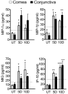
Concentrations of MIP-1α, MIP-1β, MIG, and IP-10 in the corneal epithelium and conjunctiva of untreated (UT) mice and mice with experimental dry eye for 5 (5D) and 10 (10D) days, as determined by immunobead assay. Data are expressed as the mean ± SEM. Dotted line: lowest value in the linear portion of the curve generated from the observed mean fluorescence intensities versus the observed concentrations. *P < 0.05, **P < 0.01, ***P < 0.001 vs. UT. (Adapted and modified from Yoon et al., 2007).
Figure 5
Figure 5
Representative confocal images of center of whole-mount corneas showing CD11b+ cells (green) in normal (A) and experimental dry eyes (B). (Adapted from Rashid et al., 2008).
Figure 6
Figure 6. In vivo confocal microscopy representative image (Confoscan 4, Nidek Technologies, Italy) of the cornea in a patient with DED
Numerous dendritic-like cells are demonstrated in the epithelial cell basal layer, whereas in normal age-matched controls very few of these cells are detected.
Figure 7
Figure 7. Analysis of corneal lymphangiogenesis in normal and dry eye mice
Corneas were immunostained with CD31 (green) and Lyve1 (red) antibodies. Newly-formed lymphatic vessels (CD31lo/LYVE-1+) were seen in dry eye disease (DED) corneas. IL-17 blockade significantly reduced the lymphangiogenesis compared to untreated and isotype-Ab-treated DED corneas. (Adapted from Chauhan et al., 2011).
Figure 8
Figure 8. Detection of IFN-γ-secreting Th1 and IL-17-secreting Th17 cells in draining lymph node (LN) of normal (NL) and dry eye (DE) mice
(A) ELISPOT assay for IFN-γ secretion of T cells. The results are depicted as the mean number of spots per 0.5 million responder T cells loaded ± SEM. (Adapted and modified from El Annan et al., 2009). (B) Flow cytometry analysis of Th17 cells (p = 0.026). (Adapted and modified from Chauhan et al., 2009).

References

    1. Abelson MB, Heller W, Shapiro AM, Si E, Hsu P, Bowman LM AzaSite Clinical Study Group. Clinical cure of bacterial conjunctivitis with azithromycin 1%: vehicle-controlled, double-masked clinical trial. Am J Ophthalmol. 2008;145:959–965. - PubMed
    1. Ambati BK, Patterson E, Jani P, Jenkins C, Higgins E, Singh N, Suthar T, Vira N, Smith K, Caldwell R. Soluble vascular endothelial growth factor receptor-1 contributes to the corneal antiangiogenic barrier. Br J Ophthalmol. 2007;91:505–508. - PMC - PubMed
    1. Ambati BK, Nozaki M, Singh N, Takeda A, Jani PD, Suthar T, Albuquerque RJ, Richter E, Sakurai E, Newcomb MT, Kleinman ME, Caldwell RB, Lin Q, Ogura Y, Orecchia A, Samuelson DA, Agnew DW, St Leger J, Green WR, Mahasreshti PJ, Curiel DT, Kwan D, Marsh H, Ikeda S, Leiper LJ, Collinson JM, Bogdanovich S, Khurana TS, Shibuya M, Baldwin ME, Ferrara N, Gerber HP, De Falco S, Witta J, Baffi JZ, Raisler BJ, Ambati J. Corneal avascularity is due to soluble VEGF receptor-1. Nature. 2006;443:993–997. - PMC - PubMed
    1. Amin AR, Attur MG, Thakker GD, Patel PD, Vyas PR, Patel RN, Patel IR, Abramson SB. A novel mechanism of action of tetracyclines: effects on nitric oxide syntheses. Proc Natl Acad Sci USA. 1996;93:14014–14019. - PMC - PubMed
    1. Aragona P, Bucolo C, Spinella R, Giuffrida S, Ferreri G. Systemic omega-6 essential fatty acid treatment and pge1 tear content in Sjögren’s syndrome patients. Invest Ophthalmol Vis Sci. 2005;46:4474–4479. - PubMed

Publication types

LinkOut - more resources