. 2012 Apr;122(4):1246-61.
doi: 10.1172/JCI61135. Epub 2012 Mar 19.

Radiation treatment inhibits monocyte entry into the optic nerve head and prevents neuronal damage in a mouse model of glaucoma

Affiliations

Radiation treatment inhibits monocyte entry into the optic nerve head and prevents neuronal damage in a mouse model of glaucoma

Gareth R Howell et al. J Clin Invest. 2012 Apr.

Abstract

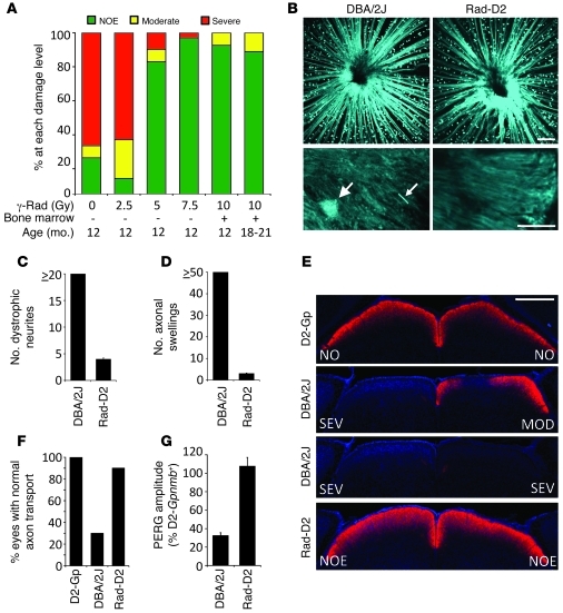
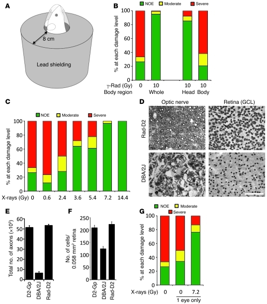
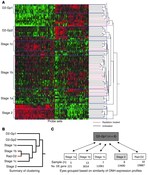
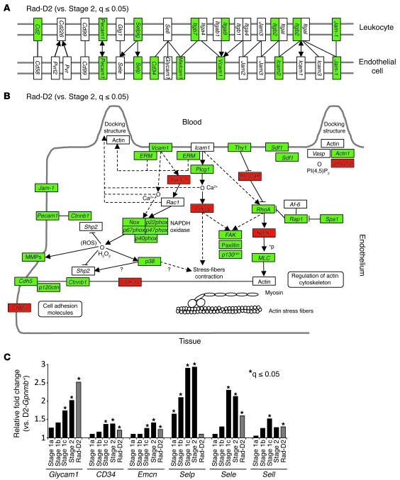
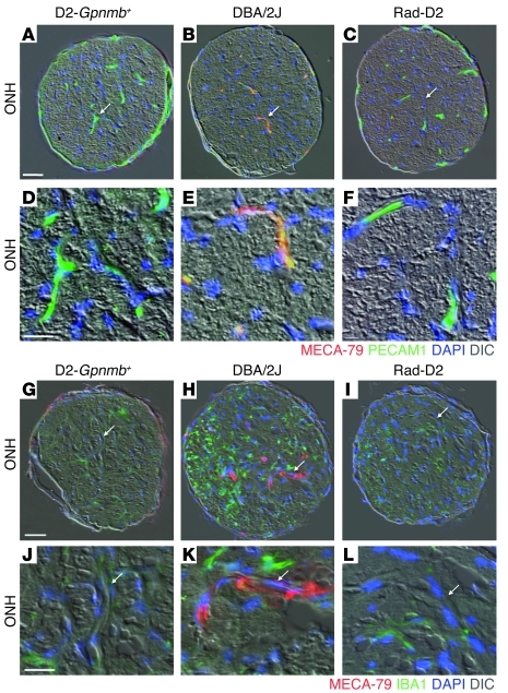
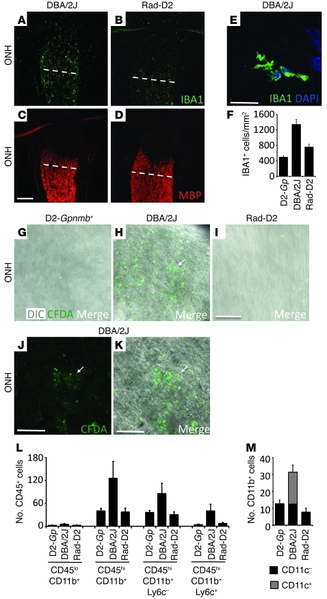
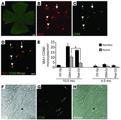
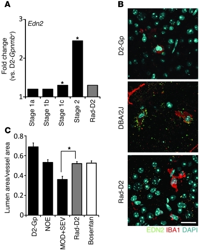
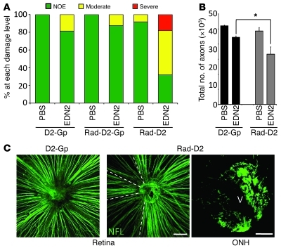
Glaucoma is a common ocular disorder that is a leading cause of blindness worldwide. It is characterized by the dysfunction and loss of retinal ganglion cells (RGCs). Although many studies have implicated various molecules in glaucoma, no mechanism has been shown to be responsible for the earliest detectable damage to RGCs and their axons in the optic nerve. Here, we show that the leukocyte transendothelial migration pathway is activated in the optic nerve head at the earliest stages of disease in an inherited mouse model of glaucoma. This resulted in proinflammatory monocytes entering the optic nerve prior to detectable neuronal damage. A 1-time x-ray treatment prevented monocyte entry and subsequent glaucomatous damage. A single x-ray treatment of an individual eye in young mice provided that eye with long-term protection from glaucoma but had no effect on the contralateral eye. Localized radiation treatment prevented detectable neuronal damage and dysfunction in treated eyes, despite the continued presence of other glaucomatous stresses and signaling pathways. Injection of endothelin-2, a damaging mediator produced by the monocytes, into irradiated eyes, combined with the other glaucomatous stresses, restored neural damage with a topography characteristic of glaucoma. Together, these data support a model of glaucomatous damage involving monocyte entry into the optic nerve.


Figures

Figure 1
Figure 1. Sublethal γ-radiation protects DBA/2J mice from glaucoma.
(A) Different doses of radiation protect from glaucoma. Most 18- to 21-month-old treated mice had no detectable optic nerve damage (NOE, n = 27). Doses of 7.5 Gy (n = 60, 12 months of age) and 5.0 Gy (n = 41) were highly protective compared to no treatment (0 Gy, n = 140) and equivalent to 10 Gy plus BMT (n = 54). 2.5 Gy (n = 35) was not protective. (BD) ONHs of DBA/2J-Thy1(CFP) and irradiated DBA/2J-Thy1(CFP) 10-month-old mice were assessed for dystrophic neurites (big arrow) and axonal swellings (little arrow) by confocal microscopy. (B) Upper panels show compressed Z stacks of DBA/2J and Rad-D2 eyes imaging from the nerve fiber layer through the glial lamina. (C) Dystophic neurites and (D) axonal swellings were greatly reduced in irradiated compared to untreated eyes (n = 6 per group). (E and F) In the majority of 10-month-old Rad-D2 eyes (18/20), anterograde axon transport to the superior colliculus (SC; assessed using a fluorescent tracer) was no different than that of controls (D2-Gpnmb+ [D2-Gp], 10/10). The majority of untreated DBA/2J mice had severe reductions of labeling in the SC (13/20). The degree of optic nerve damage in corresponding eyes is indicated (E, NO = no damage, MOD = Moderate damage, SEV = Severe damage). (G) In contrast to untreated mice, 12-month-old Rad-D2 mice had normal PERG amplitudes. Untreated had reduced PERG amplitude by 9 months of age (n = 20 each group). Scale bars: 100 μm (B, top); 20 μm (B, bottom); 500 μm (E).
Figure 2
Figure 2. Local radiation robustly protects from glaucoma in DBA/2J mice (assessed at 12 months of age).
(A) Lead shielding allowed either head-only irradiation (shown) or body-only irradiation (all of body without head). (B) Irradiation of the head robustly prevented glaucoma compared with untreated eyes (P = 1.8 × 10⁻³², treated 87/102 eyes, 85% NOE; untreated 37/140 eyes, 26% NOE). Body-only irradiation was not protective (7/34 eyes, 21% NOE). For comparison, a summary of the 0 and 10 Gy whole body data from Figure 1 is included. (CE) X-ray irradiation of just the eye protects from glaucoma. (C) X-ray doses of 7.2 and 14.4 Gy protected the vast majority of eyes from glaucoma (7.2 Gy, 58/60 eyes, 96% NOE; 14.4 Gy, 40/40 eyes, 100% NOE). 3.6 Gy and 5.4 Gy more than doubled the number of NOE eyes compared to no treatment (3.6 Gy, 32/50 eyes, 64% NOE; 5.4 Gy, 33/54 eyes, 61%). Doses of 0.6 and 2.4 Gy were not protective (0.6 Gy, 2/17 eyes, 11% NOE; 2.4 Gy, 9/32 eyes, 28%). (DF) Optic nerve and ganglion cell layer (GCL) phenotypes for 12-month-old DBA/2J and radiation-treated (7.2 Gy) DBA/2J mice. Eyes with the most common phenotype for each experimental group were assessed. Optic nerve cross-sections were stained with PPD and flat-mounted retinas were stained with cresyl violet. Scale bar: 50 μm. Protection was evident by (E) axon and (F) ganglion cell layer counts. (G) A 1-time x-ray dose of 7.2 Gy to a single eye protects from glaucoma, while the untreated fellow eye remained susceptible to glaucoma (n > 40 for each treatment class, P = 2 × 10⁻⁷).
Figure 3
Figure 3. Hierarchical clustering identified early stages of glaucoma.
(A) Glaucoma-relevant probe sets were used to cluster eyes based on similarity of ONH expression profiles. All eyes included in this study were at early stages, prior to optic nerve damage (see Methods). Four molecularly defined stages containing at least 5 DBA/2J eyes were identified (stages 1a, 1b, 1c, and 2; named for consistency with a previous study, ref. 26). Stages were ordered based on the increasing number of DE genes compared with those in the D2-Gpnmb+ control group (D2-Gp1) and based on the previous study, which also identified stage 2 and subsequent stages. Normalized intensity values for probe sets are represented as “green to black to red,” with green indicating lower normalized intensity values, and red indicating higher normalized intensity values. (B) A summary of the relationships among control groups, stages of glaucoma, and the radiation-treated group. The dendrogram shows that radiation-treated eyes (red) are most similar to eyes of stage 1b. In fact, 10 radiation-treated eyes clustered with the eyes in stage 1b (see A). This demonstrates that stresses and early molecular changes that occur in glaucoma persist in radiation-treated eyes, but the treatment prevents further progression. The sensitivity of this clustering approach is evident by the splitting of control eyes into 2 groups (D2-Gp1 and D2-Gp2, see Methods). (C) Pairwise comparisons between molecular groups and D2-Gp1. The sample number and the number of DE genes for each group are shown. Similar results were obtained when comparing glaucoma stages to D2-Gp2.
Figure 4
Figure 4. Genes involved in leukocyte transendothelial migration are expressed at lower levels in radiation-treated eyes compared with those in untreated eyes.
(A and B) Many genes in the KEGG (A) cell adhesion molecule and (B) leukocyte transendothelial migration pathways are downregulated in the Rad-D2 group compared with those in the glaucoma stage 2 group (green, significantly downregulated; red, significantly upregulated). (C) The expression levels of the major selectins and selectin ligands are generally lower in the radiation-treated group compared with those at glaucoma stages. For instance, P selectin (Selp) is upregulated by the earliest glaucoma stage but remains indistinguishable from D2-Gpnmb+ control values in radiation-protected eyes. In contrast, expression of Glycam1, a glycosylated L selectin ligand, increases in early glaucoma stages and in the radiation-treated group compared with D2-Gpnmb+ control group. *q ≤ 0.05.
Figure 5
Figure 5. L selectin ligands are activated in DBA/2J eyes but not in radiation-treated eyes.
The MECA-79 antibody binds to all known, functionally activated L selectin ligands and marks sites of chronic inflammatory signaling. (B, E, H, and K) MECA-79 bound to vessels in the ONH of DBA/2J eyes at early stages of glaucoma, prior to detectable optic nerve damage or axon loss. No MECA-79 staining was observed in the ONHs of either (C, F, I, and L) radiation-treated or (A, D, G, and J) D2-Gpnmb+controls. n = 6 eyes for each group. (H and K) MECA-79 staining coincided with an increase in staining for IBA1, a marker of microglia and monocyte-derived cells. Arrows indicate location of blood vessels, which are shown at higher magnification in the bottom panels (DF and JL). Scale bars: 50 μm (AC and GI); 20 μm (DF and JL).
Figure 6
Figure 6. Cells infiltrate into the ONHs of glaucoma-prone eyes prior to glaucomatous damage but were not detected in radiation-treated eyes.
(AF) IBA1+ cell numbers were assessed from the ONH to the myelinated region (anterior to white line) of 10.5-month-old DBA/2J, Rad-D2, and D2-Gpnmb+ control eyes by immunofluorescence. (E) An example of the counted IBA1+ cells is shown. The number of IBA1+ cells increased in untreated eyes compared with that in controls but there are significantly fewer in Rad-D2 eyes (P = 0.0002). (GK) Cell infiltration was assessed using CFDA (injected into the spleens; see Methods). CFDA+ cells had entered the optic nerves of untreated DBA/2J eyes (n = 8) but not Rad-D2 eyes (n = 6) or control eyes (n = 6). Fluorescent cells must have taken up CFDA in the spleen. H is a merged view of J (fluorescent) and K (DIC). Arrows indicate location of CFDA+ cells. (L) Flow cytometric analysis revealed that CD45hiCD11b+ monocyte numbers increased in the ONHs of untreated 10.5-month-old DBA/2J eyes compared with those in D2-Gpnmb+ controls and radiation-treated eyes (P = 0.01). CD45hiCD11b+ monocyte numbers did not increase in radiation-treated eyes compared with D2-Gpnmb+ controls (P = 0.96). CD45hi is a marker of blood-derived infiltrating cells. One-third of the CD45hiCD11b+ cells were also positive for the proinflammatory marker Ly6c+. (M) Flow cytometric analysis also shows that the CD45hi cells that infiltrate into DBA/2J eyes are CD11b+CD11c+ double-positive monocytes. This class of cell is completely absent in radiation-treated eyes (P = 0.0007, compared with untreated eyes). Scale bars: 50 μm (AC and GI); 20 μm (D and E and J and K).
Figure 7
Figure 7. Entry of cells into the retina from the blood occurs in early glaucoma but is greatly reduced in radiation-treated eyes.
(AD) The entire nerve fiber layer and ganglion cell layer of flat-mounted retinas was assessed for IBA1+CD68+ cells. CD68 is a marker of activated or responding cells (95). Cells were classed as either ramified (elongated bodies and obvious processes) or round (rounded bodies that lacked prominent processes). (BE) Images from untreated DBA/2J eyes are shown as examples. In untreated DBA/2J and radiation-treated eyes, mosaics of IBA1+CD68 microglia were present (big arrows). In untreated eyes, the number of ramified (small arrows) or round (arrowheads) IBA1+CD68+ cells increased significantly (P < 0.001 for both comparisons) compared with that in controls. In radiation-treated eyes, the number of CD68+ ramified cells also increased significantly compared with that in controls and was not significantly different than that in DBA/2J eyes (P = 0.2). However, there were significantly fewer CD68+ round cells in radiation-treated eyes (*P < 0.001). No changes were observed to IBA1+ cells in other layers of the retina (data not shown). (FH) Infiltration of cells was assessed using CFDA in 10.5-month-old mice. Both round and ramified cells that were positive for CFDA (arrows) were present in the retinas of DBA/2J eyes, indicating infiltration from the blood stream. CFDA+ cells were not detected in the retinas of radiation-treated or D2-Gpnmb+ control eyes (data not shown). Scale bars: 500 μm (A); 50 μm (BD); 10 μm (FH).
Figure 8
Figure 8. EDN2 increases in glaucoma but not in radiation-treated eyes.
(A) Edn2 expression is upregulated in glaucoma but not in radiation-treated eyes. (B) EDN2+IBA1+ cells are readily detected in retinas of untreated DBA/2J eyes but are rare in radiation-treated eyes. Scale bar: 20 μm. (C) Endothelin-2 is a potent vasoconstrictor, and the ratio of vascular lumen to overall vessel diameter was decreased in DBA/2J eyes with moderate and severe optic nerve damage (MOD+SEV) compared with that in eyes with no glaucoma (NOE, P = 0.001). This ratio was not decreased in Rad-D2 eyes compared with that in NOE eyes (P = 0.78, n > 6 in all groups). Rad-D2 eyes had very little EDN2, and vasoconstriction was prevented to the same degree as in eyes treated with an EDN receptor antagonist (Bosentan, ref. , P = 0.90). *q ≤ 0.05.
Figure 9
Figure 9. Restoring EDN2 to radiation-treated eyes induces a pattern of damage that is characteristic of glaucoma.
EDN2, injected into the vitreous of radiation-treated eyes, caused glaucoma-like RGC damage that was not observed in control eyes. (A) Most radiation-treated, EDN2-injected DBA/2J eyes had moderate and severe optic nerve damage. This was not the case for EDN2-injected untreated D2-Gp or radiation-treated D2-Gp eyes or eyes injected with vehicle (1× PBS, n > 15 each group, all age matched). D2-Gp mice do not develop high IOP and glaucoma, and so EDN2 induces the most damage in eyes that have high IOP and possibly other stresses that are not sufficient by themselves to induce glaucoma. Thus, EDN2 is a damaging factor that conspires with other stresses to induce glaucoma but is diminished in radiation-treated eyes. (B) Axon counts demonstrated a significant increase in axon loss in radiation-treated, EDN2-injected DBA/2J eyes compared with that in D2-Gpnmb+ controls (*P < 0.0001). (C) Radiation-treated, EDN2-injected DBA/2J eyes had fan-shaped patterns of RGC loss, with discrete borders in the retina and regional axon loss in the ONHs (key characteristics of glaucoma). NFL, Anti-neurofilament antibody. Fan-shaped patterns were not observed in D2-Gpnmb+ or radiation-treated D2-Gpnmb+ controls. V, vessel. Scale bars: 100 μm (retina); 50 μm (ONH).

References

    1. Whitmore AV, Libby RT, John SW. Glaucoma: thinking in new ways-a role for autonomous axonal self-destruction and other compartmentalised processes? Prog Retin Eye Res. 2005;24(6):639–662. doi: 10.1016/j.preteyeres.2005.04.004. - DOI - PubMed
    1. Quigley HA, Broman AT. The number of people with glaucoma worldwide in 2010 and 2020. Br J Ophthalmol. 2006;90(3):262–267. doi: 10.1136/bjo.2005.081224. - DOI - PMC - PubMed
    1. Quigley HA, Addicks EM. Regional differences in the structure of the lamina cribrosa and their relation to glaucomatous optic nerve damage. Arch Ophthalmol. 1981;99(1):137–143. doi: 10.1001/archopht.1981.03930010139020. - DOI - PubMed
    1. Howell GR, et al. Axons of retinal ganglion cells are insulted in the optic nerve early in DBA/2J glaucoma. J Cell Biol. 2007;179(7):1523–1537. doi: 10.1083/jcb.200706181. - DOI - PMC - PubMed
    1. May CA, Lütjen-Drecoll E. Morphology of the murine optic nerve. Invest Ophthalmol Vis Sci. 2002;43(7):2206–2212. - PubMed

Publication types

MeSH terms