

Review
doi: 10.1038/nn.3045.
Plasticity in gray and white: neuroimaging changes in brain structure during learning
Affiliations
- PMID: 22426254
- PMCID: PMC3660656
- DOI: 10.1038/nn.3045
Item in Clipboard
Review
Plasticity in gray and white: neuroimaging changes in brain structure during learning
Nat Neurosci.
.
Abstract
Human brain imaging has identified structural changes in gray and white matter that occur with learning. However, ascribing imaging measures to underlying cellular and molecular events is challenging. Here we review human neuroimaging findings of structural plasticity and then discuss cellular and molecular level changes that could underlie observed imaging effects. Greater dialog between researchers in these different fields would help to facilitate cross-talk between cellular and systems level explanations of how learning sculpts brain structure.
Figures
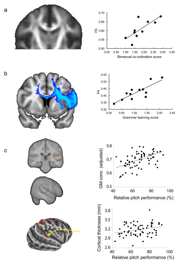
Individual differences in performance of cognitive tasks correlates with variation in gray and white matter structure of task-relevant brain areas. Figures adapted from previous publications (with permission) as follows: (A) Performance on a bimanual co-ordination task correlates with fractional anisotropy in the body of the corpus callosum, a white matter region that contains transcallosal fibers liking supplementary and cingulate motor areas. (B) Performance at acquiring the deep structure of an artificial grammar correlates with fractional anisotropy within pathways from Broca’s area, specifically in the left hemisphere. (C) gray matter concentration (top) and cortical thickness (bottom) in areas of right auditory cortex covaries with behavioral ability specifically on pitch-based tests.
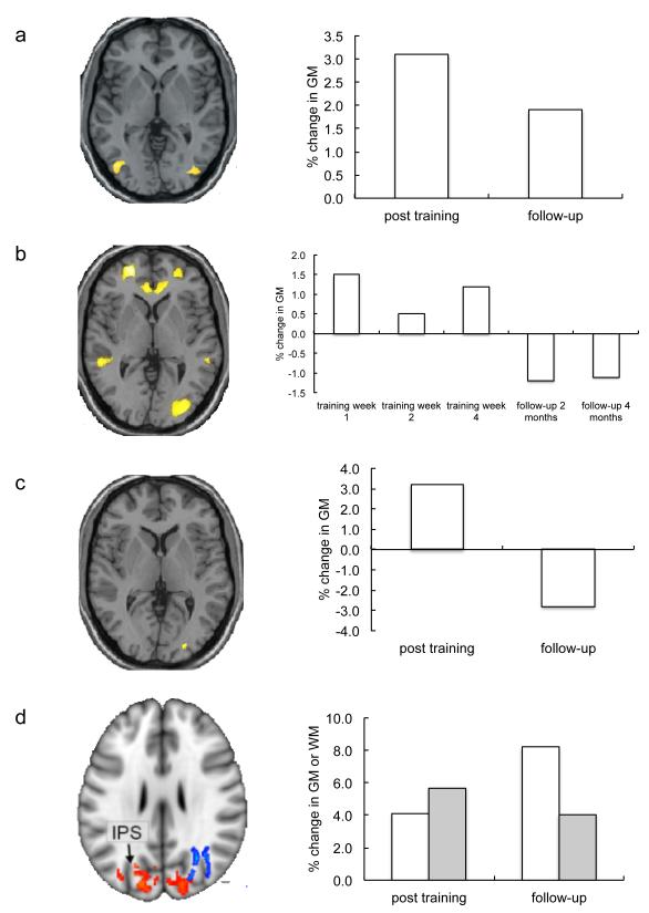
A number of studies have used learning to juggle as a paradigm to test for brain structural plasticity in healthy adults. Figures adapted from previous publications (with permission) as follows: (A) 3 months of training results in increases in gray matter density bilaterally in the visual motion area, V5. (B) Serial scans throughout the training period show that such effects are apparent as early as one week after training begins. (C) Given the same amount of training, older people learn less well on average than younger people, but those that are able to learn to juggle over the training period show similar brain structural changes. (D) Not only gray matter (red clusters on brain, white bars), but also white matter (blue clusters on brain, grey bars), shows training-related changes. Both gray matter density and white matter fractional anisotropy increase around 5% over a 6 week training period.
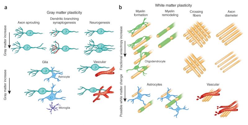
(A) Cellular events underlying changes detected by MRI during learning include axon sprouting (a), dendritic branching and synaptogenesis (b), neurogenesis (c), changes in glial number and morphology (d), and angiogenesis (e) in gray matter regions. (B) Changes in white matter include axon branching, packing density, axon diameter, fiber crossing, and the number of axons (a), myelination of unmyelinated axons (b), myelin thickness and morphology (c), changes in astrocyte morphology or number (d), and angiogenesis (e).
References
-
- He Y, Chen ZJ, Evans AC. Small-world anatomical networks in the human brain revealed by cortical thickness from MRI. Cereb Cortex. 2007;17:2407–2419. - PubMed
-
- Bermudez P, Evans AC, Lerch JP, Zatorre RJ. Neuro-anatomical correlates of musicianship as revealed by cortical thickness and voxel-based morphometry. Cerebral Cortex. 2009;19:1583–1596. - PubMed
-
- Schneider P, et al. Morphology of Heschl’s gyrus reflects enhanced activation in the auditory cortex of musicians. Nature Neuroscience. 2002;5:688–694. - PubMed
Publication types
MeSH terms
Grants and funding
LinkOut - more resources
Full Text Sources
Other Literature Sources
