

doi: 10.1038/nsmb.2260.
Structure of the activating IL-1 receptor signaling complex
Affiliations
- PMID: 22426547
- PMCID: PMC4006550
- DOI: 10.1038/nsmb.2260
Item in Clipboard
Structure of the activating IL-1 receptor signaling complex
Nat Struct Mol Biol.
.
Abstract
Interleukin-1 (IL-1)-family cytokines are mediators of innate and adaptive immunity. They exert proinflammatory effects by binding a primary receptor that recruits a receptor accessory protein to form a signaling-competent heterotrimeric complex. Here we present the crystal structure of IL-1β bound to its primary receptor IL-1RI and its receptor accessory protein IL-1RAcP, providing insight into how IL-1-type cytokines initiate signaling and revealing an evolutionary relationship with the fibroblast growth factor receptor family.
Figures
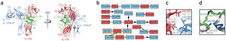
Structural features of the IL-1β signaling complex. (a) Ribbon diagram of IL-1β bound to the ectodomains of IL-1RI and IL-1RAcP in two different views, related to each other by a 130° rotation around the vertical axis. (b) Two-dimensional interaction map of the IL-1RI–IL-1RAcP interface. Amino acids are depicted as nodes. Interactions between side chains are represented by lines; interactions between side chains and backbone are depicted as arrows pointing toward the backbone. Van der Waals interactions and hydrophobic contacts are shown as solid lines, hydrogen bonds or electrostatic interactions as dashed lines. Interactions shared between the signaling and decoy receptor complex are indicated with an orange box. (c) Close-up view of the hydrophobic patch in the IL-1RI–IL-1RAcP interface that is shared between the signaling and decoy receptor complex. The decoy receptor complex is shown in gray. (d) Close-up view of selected residues in the IL-1β–IL-1RAcP interface. The hydrogen bond formed by Asn168 is depicted as a dashed line. The decoy receptor complex is shown in gray.
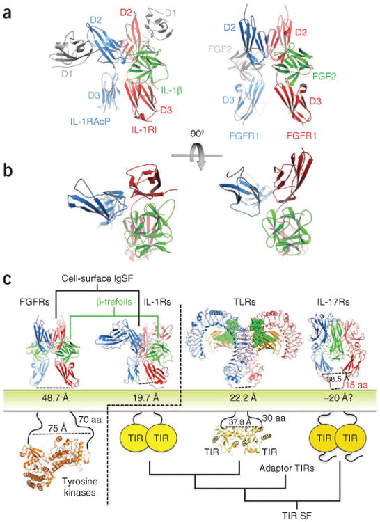
Comparison of IL-1β and FGF signaling complexes. (a) Side-by-side view of the active IL-1β–IL-1RI–IL-1RAcP heterocomplex and a representative FGF2–FGFR1 homodimeric complex. Non-equivalent N-terminal immunoglobulin domains in the IL-1 receptors, and a second bound FGF2 cytokine, are depicted in light gray. (b) Top view of the two complexes; the non-equivalent domains and second ligand have been deleted to highlight the similar interaction geometries of the three core receptors and β-trefoil ligand components. (c) Evolutionary landscape of TIR domain receptors. Structures of the FGF–FGFR complex and TIR domain receptor complexes (IL-1–IL-1RI–IL-1RAcP ternary complex, Toll-like receptor (TLR) complex and IL-17RA–IL-17F complex). Separations between receptor ectodomains as the chains enter the membrane are denoted below the respective structure. The IL-1R family architecture places the receptors at the evolutionary junction of two distinct signaling systems. FGF–FGFR homodimeric complex PDB 1FQ9 (ref. 8); FGFR kinase PDB 3CLY; TLR complex PDB 3FXI; TLR10 TIR domain PDB 2J67 (ref. 17); IL-17RA–IL-17F PDB 3JVF. SF, superfamily.
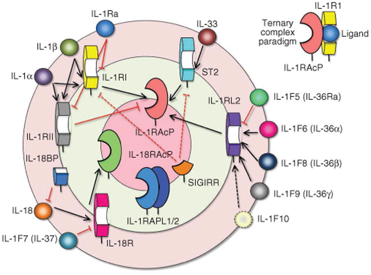
The IL-1 cytokine receptor family interaction wheel. The 11 IL-1 cytokines, are drawn on the outer rim of the wheel and fall into four functional groups that correspond to different heterodimeric receptor signaling complexes. The five primary receptors are drawn on the middle wheel and the secondary or accessory receptors on the innermost wheel (for review, see ref. 20). To form the ternary complex paradigm (top right), the primary receptors first bind their corresponding cytokine ligands and then engage the accessory receptor (which is incapable of binding cytokines by itself). In the first binding event, IL-1 cytokine ligands connect to their (often promiscuous) primary receptors (shorter black arrows), which bind secondary receptors (longer black arrows) in the final step of ternary complex assembly. Red lines (solid for known interactions, dotted for predicted) mark decoy cytokine or receptor binding that creates nonfunctional partial receptor assemblies (as in the case of IL-1Ra, which binds IL-1RI or IL-1RII but does not engage IL-1RacP) or nonfunctional complete receptor assemblies (as with IL-1RII, which binds both cytokine and IL-1RacP but lacks an intracellular signaling domain). IL-1RAPL1/2, interleukin-1 receptor accessory protein–like 1/2; ST2, growth stimulation– expressed gene 2; SIGIRR, single immunoglobulin domain–containing IL-1R–related protein.
References
-
- Sims JE, Smith DE. Nat Rev Immunol. 2010;10:89–102. - PubMed
-
- Arend WP, Palmer G, Gabay C. Immunol Rev. 2008;223:20–38. - PubMed
-
- Wang D, et al. Nat Immunol. 2010;11:905–911. - PubMed
-
- Vigers GP, Anderson LJ, Caffes P, Brandhuber BJ. Nature. 1997;386:190–194. - PubMed
-
- Murzin AG, Lesk AM, Chothia C. J Mol Biol. 1992;223:531–543. - PubMed
Publication types
MeSH terms
Substances
Associated data
- Actions
Grants and funding
LinkOut - more resources
Full Text Sources
Other Literature Sources
