. 2012 Jun;33(2):107-22.
doi: 10.1007/s10974-012-9288-7. Epub 2012 Mar 17.

Developmental regulation of MURF E3 ubiquitin ligases in skeletal muscle

Affiliations

Developmental regulation of MURF E3 ubiquitin ligases in skeletal muscle

Sue Perera et al. J Muscle Res Cell Motil. 2012 Jun.

Abstract

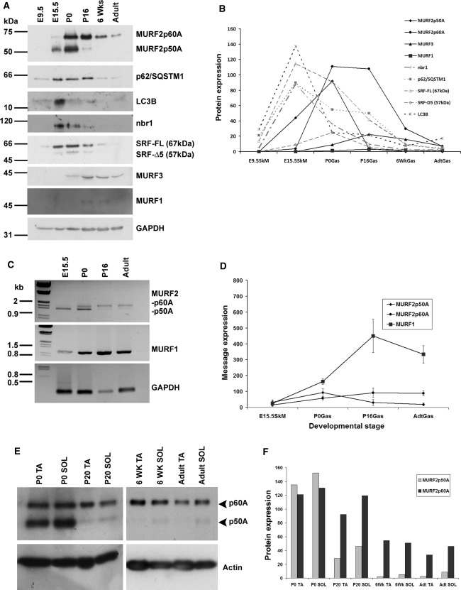
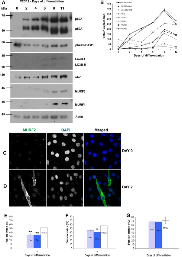
The striated muscle-specific tripartite motif (TRIM) proteins TRIM63/MURF1, TRIM55/MURF2 and TRIM54/MURF3 can function as E3 ubiquitin ligases in ubiquitin-mediated muscle protein turnover. Despite the well-characterised role of MURF1 in skeletal muscle atrophy, the dynamics of MURF isogene expression in the development and early postnatal adaptation of skeletal muscle is unknown. Here, we show that MURF2 is the isogene most highly expressed in embryonic skeletal muscle at E15.5, with the 50 kDa A isoform predominantly expressed. MURF1 and MURF3 are upregulated only postnatally. Knockdown of MURF2 p50A by isoform-specific siRNA results in delayed myogenic differentiation and myotube formation in vitro, with perturbation of the stable, glutamylated microtubule population. This underscores that MURF2 plays an important role in the earliest stages of skeletal muscle differentiation and myofibrillogenesis. During further development, there is a shift towards the 60 kDa A isoform, which dominates postnatally. Analysis of the fibre-type expression shows that MURF2 A isoforms are predominantly slow-fibre associated, whilst MURF1 is largely excluded from these fibres, and MURF3 is ubiquitously distributed in both type I and II fibres.


Figures

Fig. 1
Fig. 1
Developmental expression of MURF E3 ubiquitin ligases and related UPS/autophagy proteins in mouse skeletal muscle. a Western blot analysis of mouse skeletal muscle lysates (gastrocnemius) from embryonic (E) to postnatal (P) stages. b Plot of the densitometric analysis on Western blots normalised for GAPDH levels. c Semi-quantitative RT-PCR on cDNA isolated from mouse gastrocnemius. d Plot of semi-quantitative RT-PCR data obtained from three independent sets of experiments; error bars represent standard deviation. e Western blots showing the differential expression of MURF2 isoforms between predominantly fast (tibialis anterior––TA) and slow (soleus––SOL) muscle from P0 to adult stages. Note that p50 remains detectable postnatally only in soleus muscle
Fig. 2
Fig. 2
Fibre-type restriction of MURF2 expression. Cross-sections of gastrocnemius muscle were analysed by immunofluorescence microscopy. a, b MURF2 is ubiquitously present in all muscle fibres at P0, as detected by both the A-isoform specific HPC antibody and the p60A/B-specific HP60 antibody. c By P7, MURF2-A isoforms show slow-fibre expression. d At P7, the HP60 antibody shows that MURF2 p60A and B are more widely distributed. e, f In adult muscle, MURF2-A isoforms are again predominantly slow-fibre associated whilst HP60 staining gives a more widespread signal. Slow fibres were stained with monoclonal anti-slow myosin antibody A4.840, all myosin isoforms were visualised by the monoclonal anti- myosin antibody A1025. Scale bars 20 μm
Fig. 3
Fig. 3
MURF family members have differential fibre-type-specific expression. a MURF2-A isoforms are strongly detected in type I/slow fibres stained by the monoclonal anti-slow myosin antibody A4.840. b MURF1 appears to be excluded from the slow fibres. c MURF3 is ubiquitously present in both fast and slow fibres. Cryosectioned samples of adult soleus were stained, and imaged by confocal microscopy. Scale bars 20 μm
Fig. 4
Fig. 4
MURF expression is consistent both in vivo and in vitro and loss of MURF2 delays myogenic differentiation. a Analysis of MURFs and related UPS- and autophagy proteins in the murine C2C12 cell line as differentiation progressed. b Densitometric analysis of the Western blots normalised to GAPDH levels. c MURF2 is not detected in dividing myoblasts in vitro. d Serum withdrawal results in dramatic upregulation of MURF2-A isoforms at day 2 of myoblast differentiation. e SiRNA knockdown of MURF2 p50A or p60A results in significantly reduced fusion indexes for differentiating myoblasts. Myoblasts were considered to be differentiated if they had multiple nuclei and made cell-to-cell contacts with neighbouring cells. By day 3, degree of differentiation in cultures transfected with p50A- or p60A-specific siRNA were significantly lower than controls (p < 0.01). f By day 5, these cells were recovering and catching up with controls, however p60A-knockdown cultures still lagged behind controls (p < 0.05). g By day 7, control and siRNA-transfected myotubes possess similar degrees of differentiation. Data given as the mean percentage of transfected fused myotubes, obtained by counting >40 myoblasts from three sets of experiments. Error bars represent standard deviation. Statistical analysis was done by One-way ANOVA
Fig. 5
Fig. 5
siRNA knockdown of MURF2 p50A leads to sarcomeric disassembly in vitro. a MURF2 and α-actinin (Z-disk marker) are strongly upregulated in differentiating myoblasts at day 2 of serum withdrawal. The 2-fold magnified insert shows α-actinin appearing in the first cytoskeletal structures. Note that neighbouring, non-differentiating cells do not express MURF2 and α-actinin, only their nuclei are visible. b Knockdown of MURF2 p50A in differentiating C2C12 cells results in reduced α-actinin levels and organisation at 2 days. Scale bars 10 μm
Fig. 6
Fig. 6
MURF2 knockdown disrupts microtubule post-translational modifications in differentiating myoblasts in vitro. a, c MURF2 is strongly colocalised with the glutamylated and acetylated microtubule populations (stable) in differentiating C2C12 cells, but not with the dynamic tyrosinated fraction. Inserts in a, b: ×2 magnified regions showing colocalisation (yellow in overlay) with glutamylated and acetylated microtubules but not the tyrosinated fraction. d Knockdown of p50A leads to decreased levels and organisation of the stable, glutamylated microtubule population in myoblasts at day 2 of differentiation. The image is overexposed to show the weak residual staining for glutamylated tubulin. e In contrast, the dynamic, tyrosinated microtubule pool appears unaffected by loss of p50A. Scale bars 10 μm

References

    1. Allen DL, Leinwand LA. Postnatal myosin heavy chain isoform expression in normal mice and mice null for IIb or IId myosin heavy chains. Dev Biol. 2001;229:383–395. doi: 10.1006/dbio.2000.9974. - DOI - PubMed
    1. Bjorkoy G, Lamark T, Brech A, Outzen H, Perander M, Overvatn A, Stenmark H, Johansen T. p62/SQSTM1 forms protein aggregates degraded by autophagy and has a protective effect on huntingtin-induced cell death. J Cell Biol. 2005;171:603–614. doi: 10.1083/jcb.200507002. - DOI - PMC - PubMed
    1. Bodine SC, Latres E, Baumhueter S, Lai VK, Nunez L, Clarke BA, Poueymirou WT, Panaro FJ, Na E, Dharmarajan K, Pan ZQ, Valenzuela DM, DeChiara TM, Stitt TN, Yancopoulos GD, Glass DJ. Identification of ubiquitin ligases required for skeletal muscle atrophy. Science. 2001;294:1704–1708. doi: 10.1126/science.1065874. - DOI - PubMed
    1. Braun T, Gautel M. Transcriptional mechanisms regulating skeletal muscle differentiation, growth and homeostasis. Nat Rev Mol Cell Biol. 2011;12:349–361. doi: 10.1038/nrm3118. - DOI - PubMed
    1. Brummelkamp TR, Bernards R, Agami R. A system for stable expression of short interfering RNAs in mammalian cells. Science. 2002;296:550–553. doi: 10.1126/science.1068999. - DOI - PubMed

Publication types

MeSH terms

LinkOut - more resources