Review
. 2012 Mar 16;110(6):870-4.
doi: 10.1161/CIRCRESAHA.111.246504.

Zebrafish in the study of early cardiac development

Affiliations
Review

Zebrafish in the study of early cardiac development

Jiandong Liu et al. Circ Res. .

Abstract

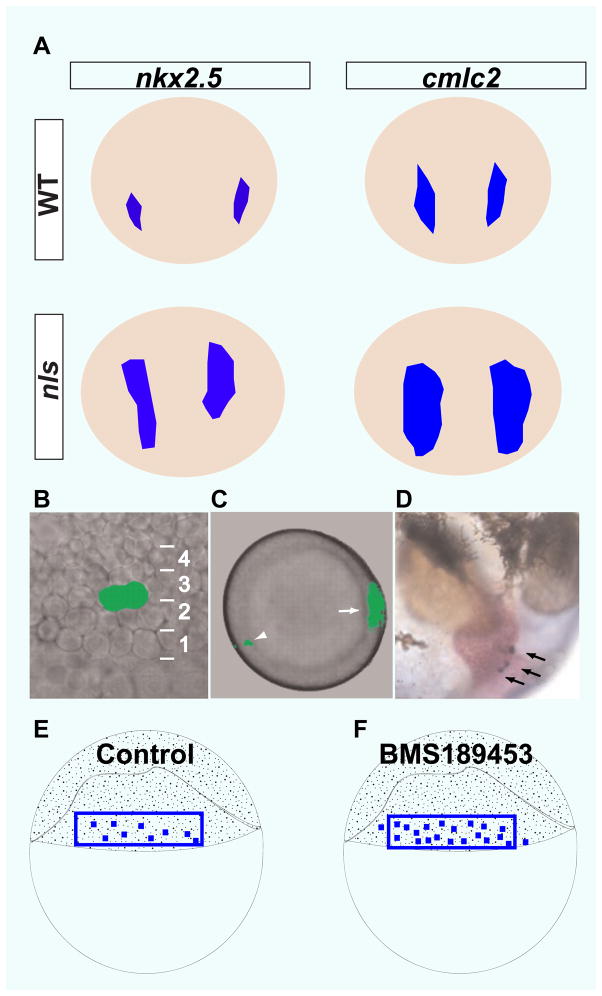
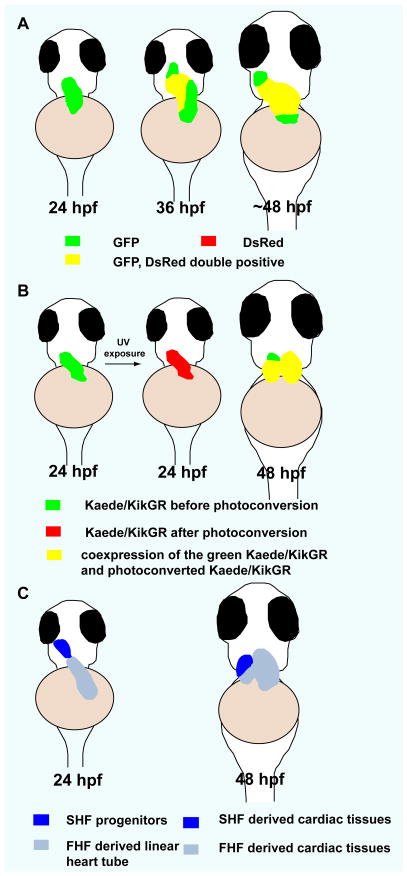
Heart development is a complex process that involves cell specification and differentiation, as well as elaborate tissue morphogenesis and remodeling, to generate a functional organ. The zebrafish has emerged as a powerful model system to unravel the basic genetic, molecular, and cellular mechanisms of cardiac development and function. We summarize and discuss recent discoveries on early cardiac specification and the identification of the second heart field in zebrafish. In addition to the inductive signals regulating cardiac specification, these studies have shown that heart development also requires a repressive mechanism imposed by retinoic acid signaling to select cardiac progenitors from a multipotent population. Another recent advance in the study of early zebrafish cardiac development is the identification of the second heart field. These studies suggest that the molecular mechanisms that regulate the second heart field development are conserved between zebrafish and other vertebrates including mammals and provide insight into the evolution of the second heart field and its derivatives.


Figures

Fig 1
Fig 1
RA signaling restricts cardiogenic potential. A) nkx2.5 and cmlc2 expression domains are expanded in aldh1a2 (aka neckless) mutant embryos compared to wildtype (6-somite stage for nkx2.5 expression and 16-somite stage for cmlc2 expression). Dorsal views, anterior to the top. B–D) Construction of blastomere fate map with caged fluorescein. B) Lateral view of two neighboring blastomeres (40% epiboly stage) in tier 3 that were labeled by activating caged fluorescein. C) Animal pole view of labeled blastomeres (arrowhead). The dorsal midline was labeled by Tg(gsc:GFP) expression (arrow). D) Descendants of the labeled blastomeres were identified in the heart (arrows, 44 hpf (hours post-fertilization)). (E–F) Schematics illustrate that more blastomeres have cardiogenic potential in BMS189453-treated embryos than in control (40% epiboly blastula stage). (adapted from Keegan BR et al., Science. 307:207–209).
Fig 2
Fig 2
Second heart field in zebrafish. A) Temporal order of cardiomyocyte differentiation: newly differentiated cardiomyocytes (green), earlier differentiated cardiomyocytes (yellow). B) Kaede/kikGR photoconversion experiments suggest that cardiomyocytes at the arterial pole of the zebrafish heart differentiate after the formation of the linear heart tube (green). C) Contribution of the SHF to zebrafish heart development. At 24 hpf, SHF progenitors are located immediately adjacent to the ventricle. At 48 hpf, SHF derived cells populate the distal segment of the ventricle as well as the outflow tract. Ventral views, anterior to the top. (adapted from de Pater E et al., Development. 136:1633–1641, Lazic and Scott, Developmental biology. 354:123–133, and Zhou Y et al., Nature. 474:645–648)

References

    1. Chien KR. Lost and found: cardiac stem cell therapy revisited. The Journal of clinical investigation. 2006;116:1838–1840. - PMC - PubMed
    1. Laugwitz KL, Moretti A, Lam J, Gruber P, Chen Y, Woodard S, Lin LZ, Cai CL, Lu MM, Reth M, Platoshyn O, Yuan JX, Evans S, Chien KR. Postnatal isl1+ cardioblasts enter fully differentiated cardiomyocyte lineages. Nature. 2005;433:647–653. - PMC - PubMed
    1. Olson EN. A genetic blueprint for growth and development of the heart. Harvey lectures. 2002;98:41–64. - PubMed
    1. Olson EN, Schneider MD. Sizing up the heart: development redux in disease. Genes & development. 2003;17:1937–1956. - PubMed
    1. Beis D, Stainier DY. In vivo cell biology: following the zebrafish trend. Trends in cell biology. 2006;16:105–112. - PubMed

Publication types