. 2012 Jun;61(6):1519-26.
doi: 10.2337/db11-0784. Epub 2012 Mar 16.

Prevention of autoimmune diabetes by ectopic pancreatic β-cell expression of interleukin-35

Affiliations

Prevention of autoimmune diabetes by ectopic pancreatic β-cell expression of interleukin-35

Maria Bettini et al. Diabetes. 2012 Jun.

Abstract

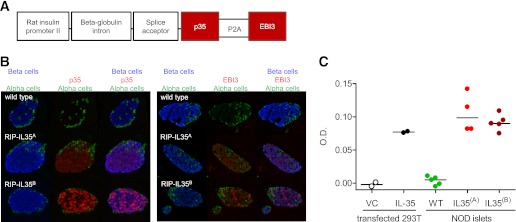
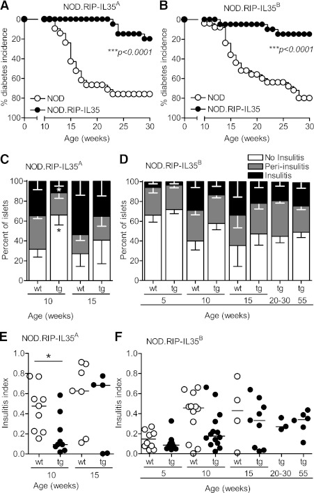
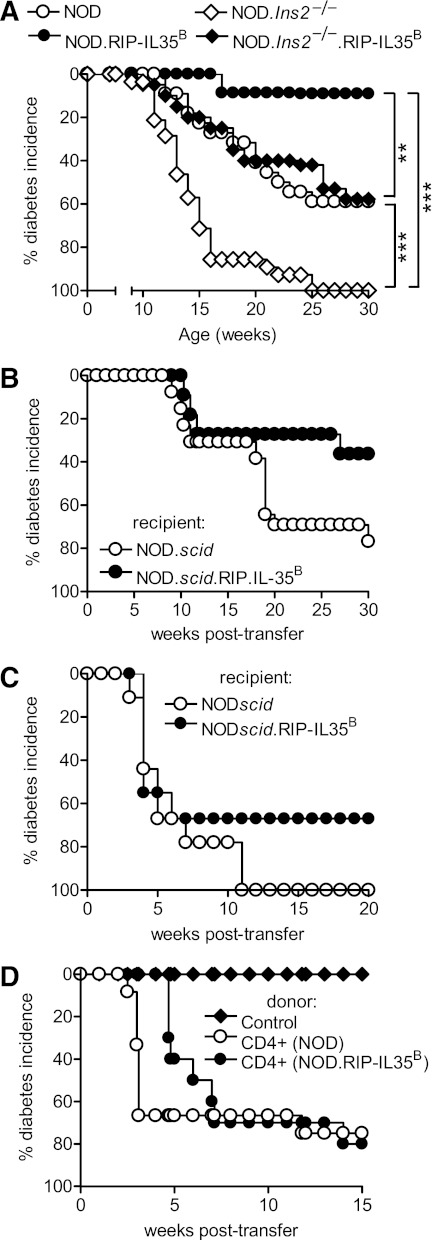
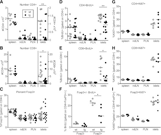
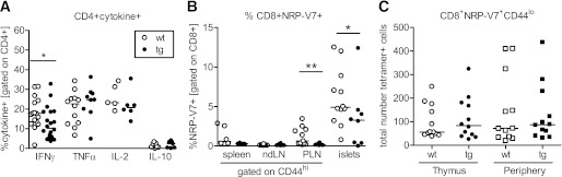
Interleukin (IL)-35 is a newly identified inhibitory cytokine used by T regulatory cells to control T cell-driven immune responses. However, the therapeutic potential of native, biologically active IL-35 has not been fully examined. Expression of the heterodimeric IL-35 cytokine was targeted to β-cells via the rat insulin promoter (RIP) II. Autoimmune diabetes, insulitis, and the infiltrating cellular populations were analyzed. Ectopic expression of IL-35 by pancreatic β-cells led to substantial, long-term protection against autoimmune diabetes, despite limited intraislet IL-35 secretion. Nonobese diabetic RIP-IL35 transgenic mice exhibited decreased islet infiltration with substantial reductions in the number of CD4(+) and CD8(+) T cells, and frequency of glucose-6-phosphatase catalytic subunit-related protein-specific CD8(+) T cells. Although there were limited alterations in cytokine expression, the reduced T-cell numbers observed coincided with diminished T-cell proliferation and G1 arrest, hallmarks of IL-35 biological activity. These data present a proof of principle that IL-35 could be used as a potent inhibitor of autoimmune diabetes and implicate its potential therapeutic utility in the treatment of type 1 diabetes.


Figures

FIG. 1.
FIG. 1.
Generation of the NOD.RIP-IL35 transgenic mouse. A: Schematic diagram of transgene construct. B: Immunofluorescent detection of p35 and EBI3 expression in the pancreatic sections of NOD.RIP-IL35A and NOD.RIP-IL35B mice. C: Detection of IL-35 in supernatant collected from cultured transgenic islets isolated from NOD.scid (green circles), NOD.scid.RIP-IL35A (red circles), and NOD.scid.RIP-IL35B (dark red circles) mice. IL-35 was measured by sandwich ELISA with a coating of primary anti-p35 and secondary biotenylated anti-EBI3. Supernatant from the 293T-cell line transfected with p35-P2A-EBI3 construct (IL-35, black circles) or vector alone (VC, white circles) was used as a control. A representative of two independent experiments is shown; each dot represents islets from a single mouse (n = 4–5). Horizontal bars represent the median. (A high-quality digital representation of this figure is available in the online issue.)
FIG. 2.
FIG. 2.
NOD.RIP-IL35 mice are protected from diabetes. NOD.RIP-IL35A (A) and NOD.RIP-IL35B (B) transgenic strains were monitored for diabetes development (n = 20–25) (***P < 0.0001, Kaplan-Meier). Insulitis and insulitis index were assessed in NOD.RIP-IL35A (black circles) (C and E) and NOD.RIP-IL35B (black circles) (D and F) transgenic strains and compared with wild-type NOD littermate controls (white circles). At least nine mice per group were analyzed at 5 and 10 weeks of age, and 3–9 mice per group were analyzed at 15–55 weeks of age (*P < 0.02, Mann-Whitney). D and F: Horizontal bars represent the median.
FIG. 3.
FIG. 3.
Transgenic expression of IL-35 confers protection against diabetes under stringent conditions. A: Ins2−/− mice were crossed with NOD.RIP-IL35B mice and monitored for diabetes onset (n = 20–28) (**P < 0.002; ***P < 0.0001, Kaplan-Meier). B: Diabetes was adoptively transferred to NOD.scid.RIP-IL35B mice by injecting 10 million splenocytes from 6- to 7-week-old female wild-type NOD mice (three separate experiments, n = 11–14). C: Diabetes was transferred by injecting 105 NY4.1 retrogenic T cells intravenously into NOD.scid-RIP-IL35B (n = 9). D: Diabetes was transferred with 5,000 CD4+ T cells sorted from the islets of 10-week-old mice in combination with CD4-depleted splenocytes from 7-week-old mice. Control group received splenocytes only (three separate experiments, n = 8–12).
FIG. 4.
FIG. 4.
Reduced T-cell infiltration and proliferation in NOD.RIP-IL35B mice. Numbers of CD4+ (A) and CD8+ (B) T cells were calculated based on flow cytometric analysis in spleens, ndLNs, PLNs, and purified islets of 10-week-old female mice (n = 7–16) (*P < 0.05; **P < 0.01, Mann-Whitney). C: Frequency of Foxp3+ cells was assessed in NOD.RIP-IL35B mice and compared with littermate controls (n = 15–17). In vivo CD4+ (D), CD8+ (E), and Foxp3+ (F) T-cell proliferation was assessed via BrdU incorporation after 4 h in vivo BrdU pulse (CD4+: n = 12–14; CD8+: n = 7–8; Foxp3+: n = 5) (*P < 0.02; **P < 0.001, Mann-Whitney). Ki67 staining of CD4+ (G), CD8+ (H), and Foxp3+ (I) T cells in organs of 10-week-old female wild-type and transgenic mice (n = 5–6). Horizontal bars represent the median.
FIG. 5.
FIG. 5.
Phenotypic analysis of islet infiltrating CD4+ and CD8+ T cells in NOD.RIP-IL35B mice. A: Cytokine production by CD4+ T cells was analyzed after 5 h in vitro restimulation with PMA and ionomycin (n = 5–16) (*P < 0.04, Mann-Whitney). B: Frequency of IGRP tetramer (NRP-V7)-positive CD8+ T cells in 10-week-old female transgenic mice and littermate controls (n = 5–12) (*P < 0.05; **P < 0.009, Mann-Whitney). C: Numbers of naive IGRP-reactive CD8+ T cells in the thymus and peripheral lymphoid organs of 5-week-old female transgenic mice and littermate controls (n = 12). Horizontal bars represent the median.

References

    1. Atkinson MA, Eisenbarth GS. Type 1 diabetes: new perspectives on disease pathogenesis and treatment. Lancet 2001;358:221–229 - PubMed
    1. Bettini M, Vignali DA. Regulatory T cells and inhibitory cytokines in autoimmunity. Curr Opin Immunol 2009;21:612–618 - PMC - PubMed
    1. Atkinson MA, Leiter EH. The NOD mouse model of type 1 diabetes: as good as it gets? Nat Med 1999;5:601–604 - PubMed
    1. Luo X, Herold KC, Miller SD. Immunotherapy of type 1 diabetes: where are we and where should we be going? Immunity 2010;32:488–499 - PMC - PubMed
    1. Chuang YP, Chu CH, Sytwu HK. Genetic manipulation of islet cells in autoimmune diabetes: from bench to bedside. Front Biosci 2008;13:6155–6169 - PubMed

Publication types

MeSH terms