. 2012 Apr 27;287(18):15024-33.
doi: 10.1074/jbc.M111.337980. Epub 2012 Mar 15.

Leucine-rich repeat and WD repeat-containing protein 1 is recruited to pericentric heterochromatin by trimethylated lysine 9 of histone H3 and maintains heterochromatin silencing

Affiliations

Leucine-rich repeat and WD repeat-containing protein 1 is recruited to pericentric heterochromatin by trimethylated lysine 9 of histone H3 and maintains heterochromatin silencing

Kui Ming Chan et al. J Biol Chem. .

Abstract

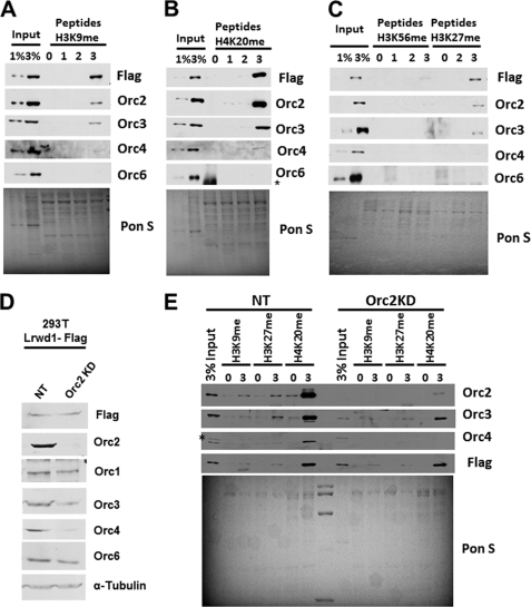
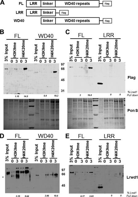
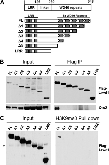
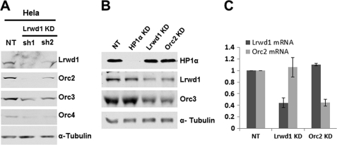
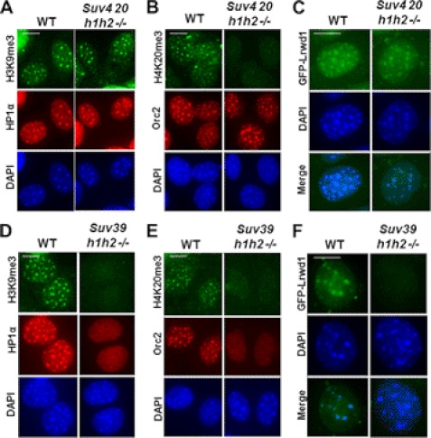
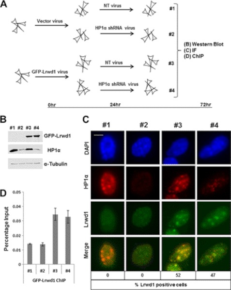
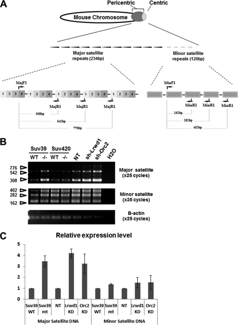
Lrwd1, a protein containing a leucine-rich repeat and a WD40 repeat domain, interacts with the origin replication complex (ORC), a protein complex involved in both initiation of DNA replication and heterochromatin silencing. Lrwd1 and ORC are known to co-purify with repressive histone marks (trimethylated lysine 9 of histone H3 (H3K9me3) and trimethylated lysine 20 of histone H4 (H4K20me3)) and localize to pericentric heterochromatin. However, how the Lrwd1 is recruited to heterochromatin and the functional significance of the localization of Lrwd1 to the heterochromatin are not known. Here, we show that Lrwd1 preferentially binds to trimethylated repressive histone marks in vitro, which is dependent on an intact WD40 domain but independent of ORC proteins. The localization of Lrwd1 and Orc2 at pericentric heterochromatin in mouse cells is lost in cells lacking H3K9me3 but not in cells lacking H4K20me3. In addition, depletion of HP1α has little impact on the localization of Lrwd1 on pericentric heterochromatin. Finally, depletion of Lrwd1 and Orc2 in mouse cells leads to increased transcription of major satellite repeats. These results indicate that the Lrwd1 is recruited to pericentric heterochromatin through binding to H3K9me3 and that the association of Lrwd1 with pericentric heterochromatin is required for heterochromatin silencing and maintenance.


Figures

FIGURE 1.
FIGURE 1.
Orc2 is not required for Lrwd1 to bind histone peptides with repressive histone marks. A–C, in vitro peptide pulldown assay was performed to assess binding of Lrwd1 and ORC proteins to peptides containing methylated histone marks. Synthetic peptides of H3K9 (A), H4K20 (B), H3K56 and H3K27 (C) with different degrees of methylation (un- (0), mono- (1), di- (2), and trimethyl (3)) were coupled to beads and used to pull down proteins in nuclear extracts prepared from 293T cells stably expressing FLAG-Lrwd1. Eluted proteins were detected by Western blotting using the indicated antibodies. Ponceau S (Pon S) staining was used to detect total proteins, which serves as loading controls. D and E, Orc2 is not required for Lrwd1 to bind H3K9me3, H3K27me3, and H4K20me3. Orc2 was depleted in 293T cells expressing FLAG-tagged Lrwd1 using shRNA, and nuclear extracts were prepared to perform the peptide pulldown assay described above using unmodified (0) and trimethylated (3) H3K9, H4K20, and H3K27 peptides. Proteins in whole cell extracts (D) as well as in pulldown (E) were detected by Western blotting using indicated antibodies. As controls, nuclear extracts were prepared from cells infected with virus for nontargeting control (NT). Each result shown is representative of three independent experiments. * indicates nonspecific band.
FIGURE 2.
FIGURE 2.
Lrwd1 WD40 domain is required and sufficient for Lrwd1 to bind repressive histone marks. A, schematic diagram shows FL Lrwd1 and two Lrwd1 mutants with either the LRR domain or WD40 repeat domain used for in vitro peptide pulldown assay. B and C, WD40 repeat domain, but not LRR domain of Lrwd1, binds H3K9me3 and H4K20me3. Nuclear extracts were prepared from 293T cells expressing FLAG-tagged full-length and truncated forms of Lrwd1, and peptide pulldown assays were performed as described in Fig. 1. D and E, recombinant full-length Lrwd1 and WD40 domain bind to methylated peptides. Recombinant full-length and truncated forms of Lrwd1 proteins were expressed and purified from Sf9 cells and used to incubate beads with unmodified or trimethylated form of H3K9 and H4K20 peptides for the peptide pulldown assay described in Fig. 1. Full-length and truncated forms of Lrwd1 recombinant proteins were detected by immunoblotting using antibodies prepared against the linker region of Lrwd1. Percentages of Lrwd1 pulldown in B and E were determined by normalization to input. Each result shown is a representative of three independent experiments. * indicates nonspecific band.
FIGURE 3.
FIGURE 3.
Intact WD40 domain of Lrwd1 is required for Lrwd1 association with Orc2 and trimethylated peptides. A, schematic diagram shows FLAG-tagged Lrwd1 and deletion mutants used for immunoprecipitation in B and in vitro peptide pulldown assays in C. B, FLAG-tagged full-length and truncated versions of Lrwd1 were expressed in 293T cells and immunoprecipitated (IP) using FLAG-M2 beads. Proteins in whole cell extracts (Input, left) and IP were analyzed by Western blotting using antibodies against Orc2 and Lrwd1. C, full-length but not truncated forms of Lrwd1 bind to H3K9me3. Nuclear extracts were prepared from cells expressing FLAG-tagged full-length or truncated versions of Lrwd1 and used for in vitro peptide pulldown assays using H3K9 and H3K9me3 peptides. Proteins in whole cell extracts (Input, left) and H3K9me3 pulldown were analyzed by Western blotting. * indicates nonspecific band.
FIGURE 4.
FIGURE 4.
Lrwd1 is required for the stability of a subset of ORC proteins in vivo. A, depletion of Lrwd1 in HeLa cells using two independent shRNA(s) results in reduced protein levels of Orc2, Orc3, and Orc4. HeLa cells were infected with two different shRNA viruses for Lrwd1 and nontargeting (NT) control. Cell lysates were prepared 72 h after infection, and ORC proteins were detected by Western blotting. B, Orc3 protein level is reduced in Lrwd1-depleted MEF cells. HP1α, Lrwd1, and Orc2 were depleted by shRNAs in MEF cells. Orc3, HP1, and Lrwd1 in whole cell extracts were analyzed by Western blotting. Note: no antibody is available to detect mouse Orc2, so Orc3 was used to monitor Orc2 protein level. C, depletion of Lrwd1 does not affect the mRNA level of Orc2. Lrwd1 or Orc2 was depleted in MEF cells as described in B. Total RNA was isolated, and the expression of Orc2 or Lrwd1 in nontargeting (NT), Lrwd1 knockdown (KD), or Orc2 knockdown (KD) cells was analyzed by real-time RT-PCR. Data and error bars indicate the mean values ± S.D. of two independent experiments.
FIGURE 5.
FIGURE 5.
H3K9me3 is required for stable association of Lrwd1 and Orc2 with pericentric heterochromatin in vivo. A–C, localization of HP1α (A), Orc2 (B), and Lrwd1 (C) at pericentric heterochromatin in wild-type and Suv420 h1/h2−/− MEF cells. D–F, localization of HP1α (D), Orc2 (E), and Lrwd1 (F) at pericentric heterochromatin compromised in Suv39 h1/h2−/− MEF cells. DAPI stains pericentric heterochromatin. Immunofluorescence using antibodies against HP1α, GFP-Lrwd1, or H3K9me3 of wild-type, Suv39 h1/h2−/−, and Suv420 h1/h2−/− MEF cells was performed as described under “Experimental Procedures.” Scale bar, 5 μm.
FIGURE 6.
FIGURE 6.
Pericentric heterochromatin localization of Lrwd1 is independent of HP1α. A, diagram shows the experimental procedures. B, Western blotting analysis of expression of GFP-Lrwd1 and HP1α in four samples from A, C, and D is shown. C, localization of Lrwd1 is unaffected in HP1α-depleted cells compared with nontargeting cells. Cells with Lrwd1 localization at pericentric heterochromatin were counted (n = 100) in three independent experiments. Scale bar, 5 μm. D, depletion of HP1α does not affect Lrwd1 binding to pericentric heterochromatin. ChIP assays using antibodies against GFP-Lrwd1 were performed, and ChIP DNA was analyzed by real-time PCR using PCR primers amplifying major satellite repeat.
FIGURE 7.
FIGURE 7.
Lrwd1 is required for silencing of major satellite repeats. A, schematic diagram showing location of major and minor satellite repeats at pericentric heterochromatin and centric heterochromatin on a mouse chromosome (upper) and organization of mouse major and minor satellite repeats and primers used to detect transcripts by RT-PCR (lower). B, RT-PCR analysis of cDNA derived from wild-type, Suv39h1h2−/−, Suv420h1h2−/− MEF cells or wild-type cells infected with virus for nontargeting control (NT), shRNA for Lrwd1 or shRNA for Orc2. One representative result of three independent experiments is shown. C, experiment performed as described in B except that transcripts of major and minor satellites were detected by real-time PCR using PCR primers described under “Experimental Procedures.” β-Actin serves as loading control. The average ± S.D. (error bars) of three independent experiments are shown. KD, knockdown.

References

    1. Grewal S. I., Jia S. (2007) Heterochromatin revisited. Nat. Rev. Genet. 8, 35–46 - PubMed
    1. Grewal S. I., Moazed D. (2003) Heterochromatin and epigenetic control of gene expression. Science 301, 798–802 - PubMed
    1. Grewal S. I., Elgin S. C. (2002) Heterochromatin: new possibilities for the inheritance of structure. Curr. Opin. Genet. Dev. 12, 178–187 - PubMed
    1. Augui S., Nora E. P., Heard E. (2011) Regulation of X-chromosome inactivation by the X-inactivation centre. Nat. Rev. Genet. 12, 429–442 - PubMed
    1. Wutz A. (2011) Gene silencing in X-chromosome inactivation: advances in understanding facultative heterochromatin formation. Nat. Rev. Genet. 12, 542–553 - PubMed

Publication types

MeSH terms