. 2012 Jul;11(7):M111.016378.
doi: 10.1074/mcp.M111.016378. Epub 2012 Mar 15.

Quantitative proteomics reveals that only a subset of the endoplasmic reticulum contributes to the phagosome

Affiliations

Quantitative proteomics reveals that only a subset of the endoplasmic reticulum contributes to the phagosome

François-Xavier Campbell-Valois et al. Mol Cell Proteomics. 2012 Jul.

Abstract

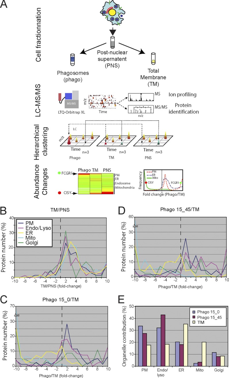
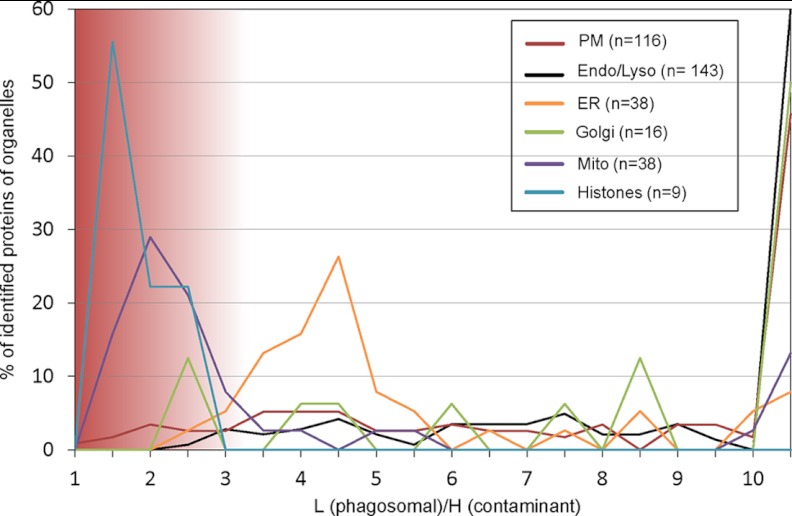
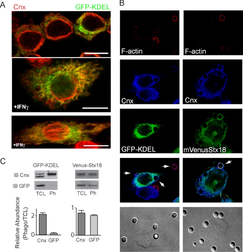
Phagosomes, by killing and degrading pathogens for antigen presentation, are organelles implicated in key aspects of innate and adaptive immunity. Although it has been well established that phagosomes consist of membranes from the plasma membrane, endosomes, and lysosomes, the notion that the endoplasmic reticulum (ER) membrane could play an important role in the formation of the phagosome is debated. However, a method to accurately estimate the contribution of potential source organelles and contaminants to the phagosome proteome has been lacking. Herein, we have developed a proteomic approach for objectively quantifying the contribution of various organelles to the early and late phagosomes by comparing these fractions to their total membrane and postnuclear supernatant of origin in the J774A.1 murine macrophage cell line. Using quantitative label-free mass spectrometry, the abundance of peptides corresponding to hundreds of proteins was estimated and attributed to one of five organelles (e.g. plasma membrane, endosomes/lysosomes, ER, Golgi, and mitochondria). These data in combination with a stable isotope labeling in cell culture method designed to detect potential contaminant sources revealed that the ER is part of the phagosomal membrane and contributes ≈ 20% of the early phagosome proteome. In addition, only a subset of ER proteins is recruited to the phagosome, suggesting that a specific subdomain(s) of the ER might be involved in phagocytosis. Western blotting and immunofluorescence substantially validated this conclusion; we were able to demonstrate that the fraction of the ER in which the ER marker GFP-KDEL accumulates is excluded from the phagosomes, whereas that containing the mVenus-Syntaxin 18 is recruited. These results highlight promising new avenues for the description of the pathogenic mechanisms used by Leishmania, Brucella, and Legionella spp., which thrive in ER-rich phagosomes.


Figures

Fig. 1.
Fig. 1.
A proteomics approach reveals the contribution of the various source organelles to the phagosome proteome. The figure shows the rationale of the MS strategy designed to assess the relative contribution of diverse organelles to the phagosome membrane. PB phagosomes (Phago15/0 or 15/45) were purified from J774.1 cells on sucrose gradients in three independent trials. In parallel, TM and PNS were isolated. A, the protein composition of all four samples was characterized by MS, and their relative abundance within the TM, PNS, Phago15/0, and Phago15/45 fractions was determined. The proteins were assigned to five organelles (PM, endosome/lysosome, ER, Golgi apparatus, and mitochondria) according to the UniProt database. Protein fold changes were then plotted according to their frequency within −10 and 10 at bins of 1. B–D, the distribution of fold change for proteins of the TM versus PNS (B), Phago15/0 versus TM (C), and Phago15/45 versus TM (D) are shown. E, estimation of the contribution of each organelle to phagosome and TM proteomes. Mean peptide intensities for all assigned proteins within an organelle were averaged, yielding an estimate of the membrane contribution of the source organelle. The intensities of all members of ER, PM, endosome/lysosome, mitochondria, and Golgi were set arbitrarily to 100%, thus ignoring other endomembrane compartments.
Fig. 2.
Fig. 2.
SILAC experiment to identify potential contaminations to the phagosome. RAW264.7 macrophages were grown in light DMEM, and phagocytosis was induced for 30 min. These cells were mixed with an equal number of cells grown in heavy labeled DMEM and lysed. Phagosomes were isolated and analyzed by quantitative MS. Subcellular localization of proteins was obtained from Uniprot, and organelles were plotted according to their ratio of light (L) to heavy (H) in bins of 0.5. A light to heavy ratio of ∼1 indicates a potential source of contamination such as mitochondria and histones, whereas most proteins associated to other organelles appear to be genuine phagosome components.
Fig. 3.
Fig. 3.
Validation of the label-free MS results by Western blotting. A, similar amounts of protein extracts from Phago15/0 and 15/45 and TM were probed with antibodies against appropriate controls and ER annotated proteins that displayed various abundance levels according to MS. B, the fold changes obtained by MS were plotted against the relative enrichment determined by densitometry of WB triplicates, yielding a linear correlation with r = 0.80. ER and other organelle annotated proteins are represented by white and gray circles, respectively. The numbers provide a link to the proteins blotted and the early and late status of phagosomes probed in each instance (supplemental Table S7).
Fig. 4.
Fig. 4.
Validation of MS results by confocal microscopy indicates that only a subdomain of the ER is recruited to the phagosome. Early PB-IgG phagosomes were formed in J774A.1 cells that were plated on fibronectin-coated cyoverslips. After fixation and permeabilization, the cells were stained for various proteins detected on the phagosome (e.g. SRP54, Stx18, and SPTLC2) and counterstained with Cnx antibody and phalloidin-BODIPY to reveal nascent phagosomes (“Experimental Procedures”). A, the data indicate that the ER proteins SRP54 and Stx18 co-localize with Cnx on the phagosome, whereas SPTLC2 does not. B, quantification of the relative co-localization in whole cells of putative phagosome markers over Cnx using the mean Pearson's coefficients obtained by the analysis of at least three representative fields for each staining reveal significant differences in the distribution of several ER markers.
Fig. 5.
Fig. 5.
mVenus-Stx18, but not GFP-KDEL, is localized to the subregion of ER implicated in phagocytosis. A, confocal microscopy of RAW264.7 stable cell line expressing GFP-KDEL in the absence (top panel) or presence (bottom two panels) of interferon-γ (IFNγ), which was used to flatten the cell to improve the spatial resolution; GFP and Cnx were detected by immunofluorescence. The bottom panel shows a flattened three-dimensional image rendered from multiple confocal sections obtained through the depth of the cell (the bar represents 10 μm). Note the discrepancy in the co-localization of GFP and Cnx, particularly in the perinuclear region and at the cell periphery. B, PB-IgG phagosomes were internalized by RAW264.7 GFP-KDEL and J774A.1 mVenus-Stx18. The cells were stained for Cnx, and F-actin was revealed by phalloidin-BODIPY to identify early phagosomes. C, WB using antibody against Cnx and GFP on phagosomes fraction obtained from the same cell line as described in B were performed to compare the recruitment of Cnx, GFP-KDEL, and mVenus-Stx18 to the phagosome fraction. TCL, total cell lysate; IB, immunoblot; Ph, phagosomes fraction.

References

    1. Jutras I., Desjardins M. (2005) Phagocytosis: At the crossroads of innate and adaptive immunity. Annu. Rev. Cell Dev. Biol. 21, 511–527 - PubMed
    1. Heine J. W., Schnaitman C. A. (1971) A method for the isolation of plasma membrane of animal cells. J. Cell Biol. 48, 703–707 - PMC - PubMed
    1. Pitt A., Mayorga L. S., Stahl P. D., Schwartz A. L. (1992) Alterations in the protein composition of maturing phagosomes. J. Clin. Invest. 90, 1978–1983 - PMC - PubMed
    1. Bajno L., Peng X. R., Schreiber A. D., Moore H. P., Trimble W. S., Grinstein S. (2000) Focal exocytosis of VAMP3-containing vesicles at sites of phagosome formation. J. Cell Biol. 149, 697–706 - PMC - PubMed
    1. Braun V., Fraisier V., Raposo G., Hurbain I., Sibarita J. B., Chavrier P., Galli T., Niedergang F. (2004) TI-VAMP/VAMP7 is required for optimal phagocytosis of opsonised particles in macrophages. EMBO J. 23, 4166–4176 - PMC - PubMed

Publication types

MeSH terms

LinkOut - more resources