Comparative Study
. 2012;7(3):e30679.
doi: 10.1371/journal.pone.0030679. Epub 2012 Mar 9.

The majority of microRNAs detectable in serum and saliva is concentrated in exosomes

Affiliations
Comparative Study

The majority of microRNAs detectable in serum and saliva is concentrated in exosomes

Alessia Gallo et al. PLoS One. 2012.

Abstract

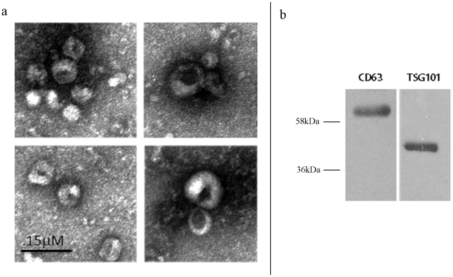
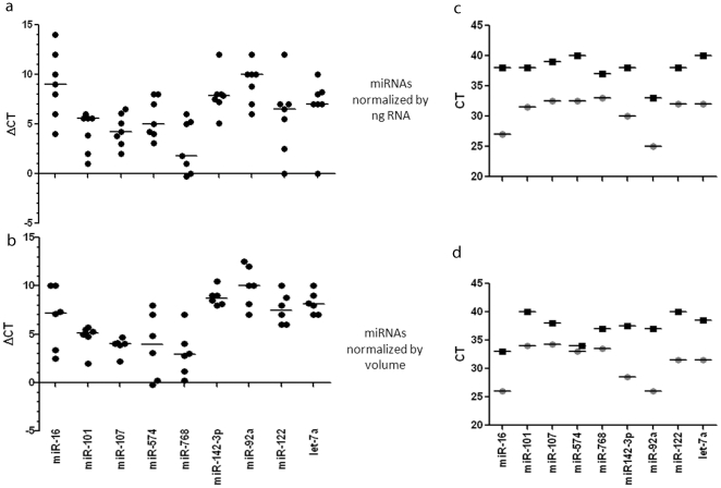
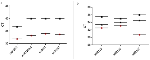
There is an increasing interest in using microRNAs (miRNA) as biomarkers in autoimmune diseases. They are easily accessible in many body fluids but it is controversial if they are circulating freely or are encapsulated in microvesicles, particularly exosomes. We investigated if the majority of miRNas in serum and saliva are free-circulating or concentrated in exosomes. Exosomes were isolated by ultracentrifugation from fresh and frozen human serum and saliva. The amount of selected miRNAs extracted from the exosomal pellet and the exosome-depleted serum and saliva was compared by quantitative RT-PCR. Some miRNAs tested are ubiquitously expressed, others were previously reported as biomarkers. We included miRNAs previously reported to be free circulating and some thought to be exosome specific. The purity of exosome fraction was confirmed by electronmicroscopy and western blot. The concentration of miRNAs was consistently higher in the exosome pellet compared to the exosome-depleted supernatant. We obtained the same results using an equal volume or equal amount of total RNA as input of the RT-qPCR. The concentration of miRNA in whole, unfractionated serum, was between the exosomal pellet and the exosome-depleted supernatant. Selected miRNAs, which were detectable in exosomes, were undetectable in whole serum and the exosome-depleted supernantant. Exosome isolation improves the sensitivity of miRNA amplification from human biologic fluids. Exosomal miRNA should be the starting point for early biomarker studies to reduce the probability of false negative results involving low abundance miRNAs that may be missed by using unfractionated serum or saliva.


Conflict of interest statement

Competing Interests: The authors have declared that no competing interests exist.

Figures

Figure 1
Figure 1. Confirmation that the ultracentrifugation pellet contains exosomes.
a Electron microscopy of the ultracentrifugation pellet from serum shows the characteristic spherical shape and size (50–100 nm) of exosomes b. Western blot shows strong staining of the ultracentrifugation pellet with the exosomal membrane markers anti cd63 and TSG101.
Figure 2
Figure 2. Serum miRNAs are predominantly in exosomes.
The relative expression of microRNAs extracted from exosome-depleted supernatant or exosomes was expressed as the difference in threshold cycle number between the exosome depleted supernatant and the exosome pellet (ΔCt: Ct supernatant – Ct exosome pellet). A 1 unit difference in ΔCT represents a two-fold difference in the amount of input miR. Positive numbers show higher concentrations in the exosomes whereas negative numbers indicate higher concentrations in the exosome-depleted supernatant. The absolute difference between the two can be calculated as 2ΔCT. The RNA included in the qPCR reaction was normalized to either equal amount (a) or equal volume (b) of RNA. Panels (c) and (d) show the absolute differences in microRNA levels amplified from serum exosome pellet (○) and exosome-depleted supernatant (□).
Figure 3
Figure 3. Salivary miRNAs are predominantly in exosomes.
(a) Absolute differences in microRNA levels amplified from saliva exosome pellet (○) and exosome-depleted supernatant (□). 1 unit increase in CT represent a two-fold lower amount of miRNA. miRNAs requiring 35 cycles or more are considered undetectable. (b) Graph showing the average of three samples in microRNA levels amplified from serum exosome pellet (○) and exosome-depleted supernatant (□) and whole serum (▵). Ct value for the whole serum samples was consistently lower (by at least 2 cycles) than the supernatant and at least 1.5 cycles higher than the exosome pellet samples.

References

    1. Alevizos I, Illei GG. MicroRNAs as biomarkers in rheumatic diseases. Nat Rev Rheumatol. 2010;6:391–8. - PMC - PubMed
    1. Kroh EM, Parkin RK, Mitchell PS, Tewari M. Analysis of circulating microRNA biomarkers in plasma and serum using quantitative reverse transcription-PCR (qRT-PCR). Methods. 2010;50:298–301. - PMC - PubMed
    1. Mitchell PS, Parkin RK, Kroh EM, Fritz BR, Wyman SK, et al. Circulating microRNAs as stable blood-based markers for cancer detection. Proc Natl Acad Sci U S A. 2008;105:10513–10518. - PMC - PubMed
    1. Heijnen HF, Schiel AE, Fijnheer R, Geuze HJ, Sixma JJ. Activated platelets release two types of membrane vesicles: microvesicles by surface shedding and exosomes derived from exocytosis of multivesicular bodies and alpha-granules. Blood. 1999;94:3791–3799. - PubMed
    1. McLellan AD. Exosome release by primary B cells. Crit Rev Immunol. 2009;29(3):203–17. - PubMed

Publication types