. 2012;7(3):e32241.
doi: 10.1371/journal.pone.0032241. Epub 2012 Mar 13.

Non-enzymatic decomposition of collagen fibers by a biglycan antibody and a plausible mechanism for rheumatoid arthritis

Affiliations

Non-enzymatic decomposition of collagen fibers by a biglycan antibody and a plausible mechanism for rheumatoid arthritis

Olga Antipova et al. PLoS One. 2012.

Abstract

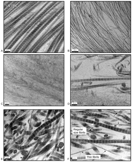
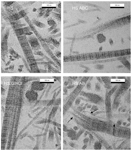
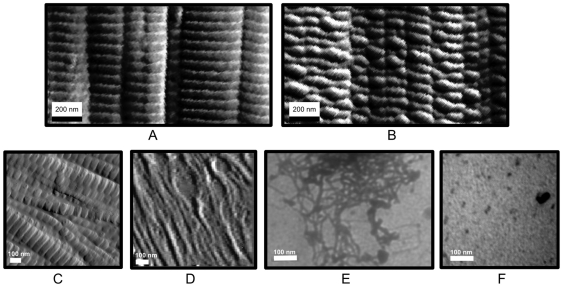
Rheumatoid arthritis (RA) is a systemic autoimmune inflammatory and destructive joint disorder that affects tens of millions of people worldwide. Normal healthy joints maintain a balance between the synthesis of extracellular matrix (ECM) molecules and the proteolytic degradation of damaged ones. In the case of RA, this balance is shifted toward matrix destruction due to increased production of cleavage enzymes and the presence of (autoimmune) immunoglobulins resulting from an inflammation induced immune response. Herein we demonstrate that a polyclonal antibody against the proteoglycan biglycan (BG) causes tissue destruction that may be analogous to that of RA affected tissues. The effect of the antibody is more potent than harsh chemical and/or enzymatic treatments designed to mimic arthritis-like fibril de-polymerization. In RA cases, the immune response to inflammation causes synovial fibroblasts, monocytes and macrophages to produce cytokines and secrete matrix remodeling enzymes, whereas B cells are stimulated to produce immunoglobulins. The specific antigen that causes the RA immune response has not yet been identified, although possible candidates have been proposed, including collagen types I and II, and proteoglycans (PG's) such as biglycan. We speculate that the initiation of RA associated tissue destruction in vivo may involve a similar non-enzymatic decomposition of collagen fibrils via the immunoglobulins themselves that we observe here ex vivo.


Conflict of interest statement

Competing Interests: Author Joseph Orgel is a section editor and member of the editorial board of PLoS ONE. The Journal had no part or influence in the research reported or in the writing of the originally submitted manuscript. A patent application has been made that pertains to data reported in this manuscript. #12/419,689: “THIN FIBRIL COLLAGEN MATERIAL AND METHOD FOR MAKING THIN FIBRIL COLLAGEN FROM NATIVE COLLAGEN FIBERS.” This does not alter the authors' adherence to all the PLoS ONE policies on sharing data and materials.

Figures

Figure 1
Figure 1. Decomposition of collagen type II fibrils in lamprey notochord viewed with TEM (A–D) and bovine articular cartilage (E and F).
A) Native (prior to fixing) type II collagen fibrils, incubated in TBS as control for fibril samples shown in B–D. Average fibril size is around 35 nm. B) Collagen type II fibrils following short incubation with anti-biglycan antibody. Fibril diameter is 10–15 nm. C) Collagen type II fibrils following incubation in GHCl. Although severely disrupted, the fibril decomposition appears less complete than that of the antibody incubation (B). D) Collagen type II sample following mechanical disruption. Disruption of native fibril structure is highly localized, with large sections still intact. E) Native bovine articular cartilage (prior to fixing and staining for TEM). F) Bovine articular cartilage post 1 hour treatment with anti-biglycan. Black arrows point to thin-fibrils, white arrows point to normal sized thick-fibrils.
Figure 2
Figure 2. Type II collagen fibrils decomposed into their basic aggregates (viewed via X-ray diffraction and TEM).
Some parts of the antibody treated samples maintain a loose alignment of the thin-fibrils allowing them to be analyzed with small angle X-ray diffraction (A), and insert B. An 11 and 4.5 nm packing function are apparent, which appear to correspond to the approximate diameter of the thin-fibrils (insert of C) and microfibrils (D). Native thick fibrils are shown in C as a comparison to the decomposition product (thin-fibrils).
Figure 3
Figure 3. Model of antibody action on type II collagen fibrils.
Coordinate models of the biglycan-type II collagen fibril complex based on the decoron-type I collagen fibril structures published recently (1) are shown with a model Fab (green) unit attaching to the biglycan (blue) epitope (colored red, A and ‘top’ view B). Because the epitope is located within a solvent filled channel of the collagen fibril , there is room for loops of the fab to dock with it, but its close proximately to the fibril-PG hydrogen bonding network located between the collagen fibril surface and the concave side of the PG-core proteins structure (1) may disrupt the positive interactions and dislodge the core protein from the fibril. Leading to the debundling of thick-fibrils into their constitutive thin-fibrils (C).
Figure 4
Figure 4. TEM images of human articular cartilage preparations.
A) Section of native human articular cartilage, incubated in TBS, that has collagen type II fibrils of regular 30–50 nm diameter (control for samples B–D). B) Section of human articular cartilage treated with ABC lyase for 24 h with some thin fibrils of collagen type II. C) Section of human articular cartilage treated with Guanidine hydrochloride for 24 h with presence of thin 10–15 nm fibrils and normal thick fibrils (fibril bundles). D) Section of human articular cartilage, treated with anti-biglycan antibody for 24 h, shows some collagen type II thin fibrils as well as fibrils of regular 30–50 nm diameter. Arrows point to decomposing fibrils.
Figure 5
Figure 5. AFM and TEM images native and Ab treated rat tail tendon and lamprey notochord treated samples.
A) Control AFM data of type I collagen fibrils: native type I fibrils. B) Control AFM data of type I collagen fibrils: Anti- PG core protein antibody conjugated with 30 nm particles attached to type I fibrils. Note that the fibrils are intact and that the gold particles are clearly discernible as densely packed globules. C) AFM image of native lamprey type II fibrils. D) AFM image of lamprey type II fibrils after treatment with gold particle conjugated anti- biglycan antibodies as in B. Compare with B. No gold particles are visible, whilst in the type I collagen control, they are clearly imaged. E) Unstained TEM of lamprey derived collagen fibrils following anti-biglycan treatment. The antibodies in this preparation were conjugated to 10 nm gold particles and were still able to decompose the thick fibrils into thin-fibrils, but no gold particles are visible after the preparation is washed in TBS. F) Unstained TEM of biglycan aggregates attached to gold particle conjugated antibodies. Gold particles are clearly visible as dense black spots that do not appear in E.

References

    1. Scott JE. Proteodermatan and proteokeratan sulfate (decorin, lumican/fibromodulin) proteins are horseshoe shaped. Implications for their interactions with collagen. Biochemistry. 1996;35:8795–8799. - PubMed
    1. Ameye L, Aria D, Jepsen K, Oldberg A, Xu T, et al. Abnormal collagen fibrils in tendons of biglycan/fibromodulin-deficient mice lead to gait impairment, ectopic ossification, and osteoarthritis. FASEB Journal. 2002;16:673–680. - PubMed
    1. Robinson PS, Huang T-F, Kazam E, Iozzo RV, Birk DE, et al. Influence of Decorin and Biglycan on Mechanical Properties of Multiple Tendons in Knockout Mice. J Biomech Eng. 2005;127:181–185. doi: 10.1115/1.1835363. - DOI - PubMed
    1. Orgel JPRO, Eid A, Antipova O, Bella J, Scott JE. Decorin Core Protein (Decoron) Shape Complements Collagen Fibril Surface Structure and Mediates Its Binding. PLoS ONE. 2009;4:e7028. doi: 10.1371/journal.pone.0007028. - DOI - PMC - PubMed
    1. Furukawa T, Ito K, Nuka S, Hashimoto J, Takei H, et al. Absence of Biglycan Accelerates the Degenerative Process in Mouse Intervertebral Disc. Spine. 2009;34:E911–E917. doi: 10.1097/BRS.0b013e3181b7c7ec. - DOI - PMC - PubMed

Publication types