. 2012 Jun 27;134(25):10469-77.
doi: 10.1021/ja301017y. Epub 2012 Mar 29.

In vitro selection of highly modified cyclic peptides that act as tight binding inhibitors

Affiliations
Free PMC article

In vitro selection of highly modified cyclic peptides that act as tight binding inhibitors

Yollete V Guillen Schlippe et al. J Am Chem Soc. .
Free PMC article

Abstract

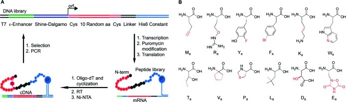
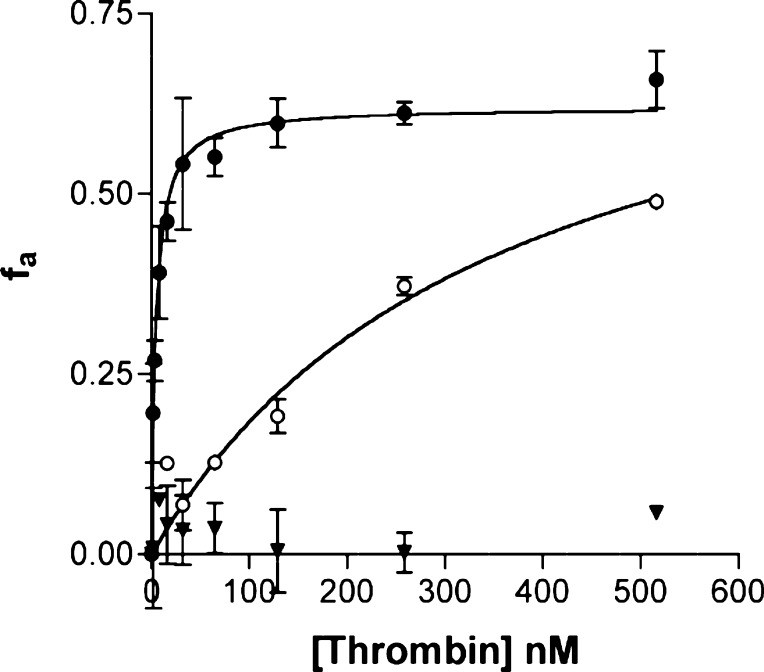
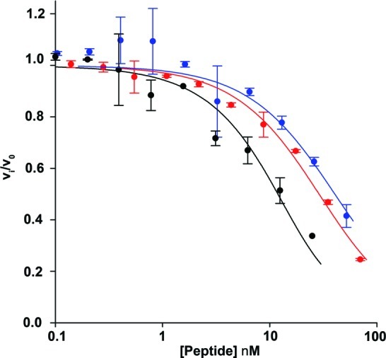
There is a great demand for the discovery of new therapeutic molecules that combine the high specificity and affinity of biologic drugs with the bioavailability and lower cost of small molecules. Small, natural-product-like peptides hold great promise in bridging this gap; however, access to libraries of these compounds has been a limitation. Since ribosomal peptides may be subjected to in vitro selection techniques, the generation of extremely large libraries (>10(13)) of highly modified macrocyclic peptides may provide a powerful alternative for the generation and selection of new useful bioactive molecules. Moreover, the incorporation of many non-proteinogenic amino acids into ribosomal peptides in conjunction with macrocyclization should enhance the drug-like features of these libraries. Here we show that mRNA-display, a technique that allows the in vitro selection of peptides, can be applied to the evolution of macrocyclic peptides that contain a majority of unnatural amino acids. We describe the isolation and characterization of two such unnatural cyclic peptides that bind the protease thrombin with low nanomolar affinity, and we show that the unnatural residues in these peptides are essential for the observed high-affinity binding. We demonstrate that the selected peptides are tight-binding inhibitors of thrombin, with K(i)(app) values in the low nanomolar range. The ability to evolve highly modified macrocyclic peptides in the laboratory is the first crucial step toward the facile generation of useful molecular reagents and therapeutic lead molecules that combine the advantageous features of biologics with those of small-molecule drugs.


Figures

Figure 1
Figure 1
In vitro selection of macrocyclic peptides using mRNA-display. (A) General scheme for the selection and amplification of cyclic peptides. The DNA library encodes peptides with 10 random amino acids flanked by two Cys residues. Following transcription and cross-linking of the RNA to a 3′-puromycin oligonucleotide, the library was translated in a completely reconstituted translation system with either all natural or replacing 12 natural amino acids with 12 unnatural amino acids to form two separate libraries of mRNA-peptide fusions. The 12 unnatural amino acids used are shown in panel B. Fusions were immobilized on an oligo-dT column and cyclized via bis-alkylation of the Cys residues, reverse transcribed (RT), and purified on a Ni-NTA resin to yield ∼2 × 10¹³ cyclic peptides. Each library was incubated separately with biotinylated thrombin in solution. Complexes were captured on streptavidin beads, and unbound material was washed away. Active mRNA–peptide fusions were eluted with a 2-fold excess of hirudin to thrombin, followed by RT-PCR amplification to generate the input material for the next round of selection/amplification. (B) Unnatural amino acids used in the unnatural peptide selection. Differences between the natural and unnatural residues are highlighted in red. The single-letter code refers to the corresponding natural amino acid that was replaced by the unnatural amino acid, with a subscript for analogue. The natural amino acids used were C, A, G, S, I, N, Q, and H.
Figure 2
Figure 2
Optimizing translation with unnatural amino acids. MALDI-TOF spectra of in vitro translation reactions with unoptimized and optimized concentrations of unnatural amino acids. (A) Reaction directed by mRNA encoding the sequence MaCVaFaGNRaGTaQPaFaCGSGSLaGHHHHHHRaLa (unnatural amino acids are labeled with the subscript a) shows several minor incorrect peaks and a major incorrect peak (×) at 3124.5 Da (most likely corresponding to −L truncation; exp −127.1 Da, obs −127). The peak at 3251.5 Da corresponds to the correct product (expected mass of 3251.4 Da). (B) In vitro translation of the same templates after optimization of the amino acid concentrations, showing improved incorporation. (C) Unoptimized in vitro translation reaction directed by mRNA encoding the sequence MaCEaFaFaDaKaKaILaAPaCGSGSLaGHHHHHHRaLa shows several minor undesired peaks and a major incorrect peak (×) at 3249.7 Da (most likely corresponding to −L truncation). The peak at 3377.9 Da corresponds to the correct product. (D) In vitro translation of the same template after optimization of amino acid concentrations, showing improved incorporation.
Figure 3
Figure 3
Selection progress and sequences of selected peptides. (A) The fraction of 35S-labeled peptide that bound to thrombin and eluted with hirudin at each round of selection is shown. Starting in round four, complexes were washed more rigorously, and in round seven the selection pressure was increased further by only amplifying mRNA–peptide fusions that remained bound after 1 h of incubation with hirudin and were subsequently eluted. Red and black bars show the progress of the unnatural and natural peptide selections, respectively. (B) Sequences of unnatural peptides after round 10. Unnatural peptide sequences U2 and U1 are labeled. Unnatural amino acids are highlighted in red, and the Cys residues used for cyclization are shown in blue; residues following the second Cys are not shown. (C) Sequence of the natural peptide winner N1 after round 10.
Figure 4
Figure 4
Verification of sequence of unnatural peptide U1. (A) Cyclization of unnatural peptide U1. Unnatural amino acids are shown in red. Cys residues used for cyclization are shown in blue. (B) LC-MS confirms formation of full-length cyclic unnatural peptide U1 (calcd [M]4+m/z = 791.62226 (4.4 ppm), [M]5+m/z = 633.49937 (0.9 ppm), [M]6+m/z = 528.08411 (0.5 ppm) (A.U., arbitrary units). (C) Peptide lacking the His6-tag was translated and the two Cys residues modified with iodoacetamide to prevent disulfide formation. LC-MS/MS analysis confirms site-specific incorporation of each unnatural amino acid into peptide U1 in the correct order. Predicted and observed ions are summarized in Tables S3–S5.
Figure 5
Figure 5
Binding of selection winners to thrombin. Binding curves for unnatural cyclic peptide U1 (closed circles), unnatural linear peptide U1 (open circles), and cyclic peptide U1 translated with natural amino acids (closed triangles). 35S-Cys-labeled peptides were incubated with varying concentrations of thrombin for 1 h. Cyclization via disulfide formation of the linear unnatural peptide U1 was prevented by the addition of 0.2 mM TCEP in the binding buffer. Bound and free peptides were separated using a 30 kDa MW cutoff spin-filter. The fraction of bound peptide fa (see Materials and Methods) was plotted against the concentration of thrombin and fit to a simple hyperbola to obtain the Kd values.
Figure 6
Figure 6
Inhibition of thrombin activity. Inhibition curves for unnatural cyclic peptide U1 (red), unnatural cyclic peptide U2 (blue), and natural cyclic peptide N1 (black). Thrombin was preincubated with varying concentrations of 35S-Cys labeled peptides for 1 h. The reaction was started by the addition of a 10mer peptide substrate (AnaSpec), and the increase in fluorescence was monitored for 1 h. Initial rates were determined, and the fraction of remaining enzymatic activity was plotted against the concentration of peptide and fit to the Morrison equation for tight-binding inhibitors to obtain Kiapp. All experiments were done at least in duplicate.

References

    1. Verdine G. L.; Walensky L. D. Clin. Cancer Res. 2007, 13, 7264. - PubMed
    1. Reichert J. M.; Rosensweig C. J.; Faden L. B.; Dewitz M. C. Nat. Biotechnol. 2005, 23, 1073. - PubMed
    1. Malik N. N. Nat. Rev. Clin. Oncol. 2009, 6, 550. - PubMed
    1. Lipinski C. A. J. Pharmacol. Toxicol. Methods 2000, 44, 235. - PubMed
    1. Swinney D. C.; Anthony J. Nat. Rev. Drug Discov. 2011, 10, 507. - PubMed

Publication types

MeSH terms