Review
. 2012 Apr;22(2):241-7.
doi: 10.1016/j.sbi.2012.02.006. Epub 2012 Mar 17.

Helical assembly in the death domain (DD) superfamily

Affiliations
Review

Helical assembly in the death domain (DD) superfamily

Ryan Ferrao et al. Curr Opin Struct Biol. 2012 Apr.

Abstract

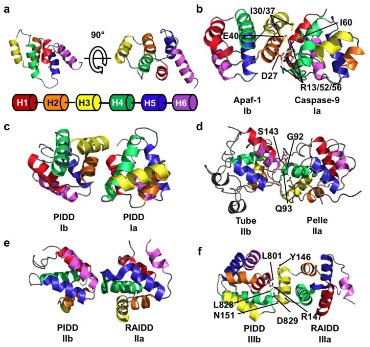
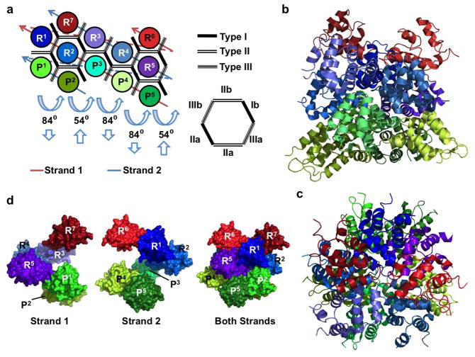
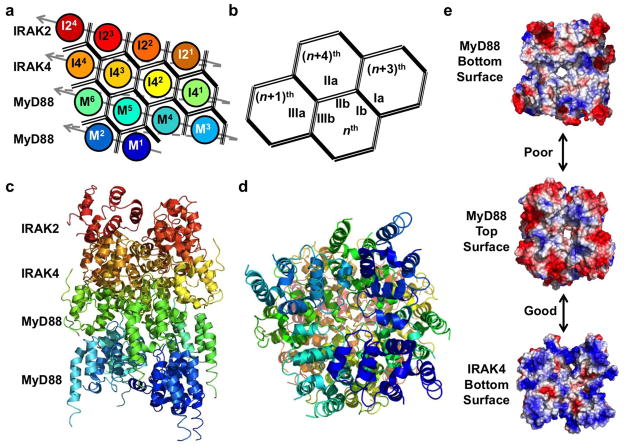
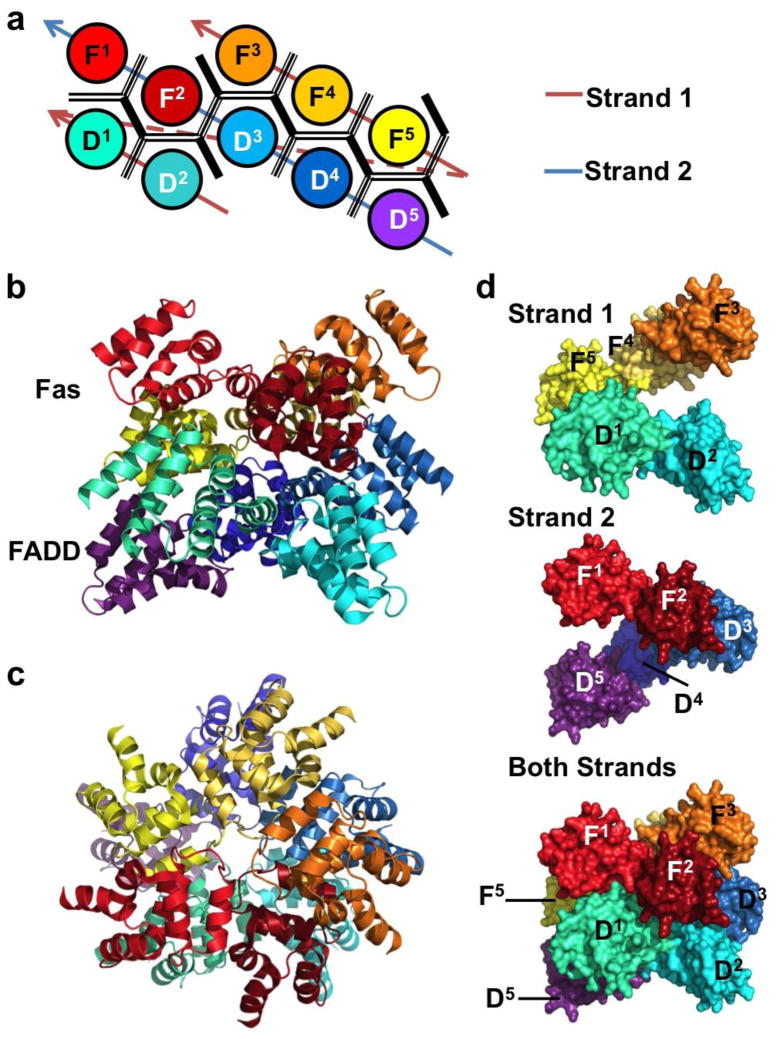
Death domain (DD) superfamily members play a central role in apoptotic and inflammatory signaling through formation of oligomeric molecular scaffolds. These scaffolds promote the activation of proinflammatory and apoptotic initiator caspases, as well as Ser/Thr kinases. Interactions between DDs are facilitated by a conserved set of interaction surfaces, type I, type II, and type III. Recently structural information on a ternary complex containing the DDs of MyD88, IRAK4, and IRAK2 and a binary complex containing Fas and FADD DDs has become available. This review will focus on how the three DD interaction surfaces cooperate to facilitate the assembly of these oligomeric signaling complexes.


Figures

Figure 1
Figure 1
Three types of asymmetric interactions mediate homotypic DD binding. (a) NMR structure of the DD of Fas, showing the six helical bundle domain architecture of the DD superfamily. Coloring scheme is shown below the structure and used throughout the figure. (b) Crystal structure of the heterodimer between the CARD domain of Apaf-1 and Caspase-9. In this type I interaction, negatively charged residues from H2 and H3 (interface Ib) of Apaf-1 interact with positively charged H1 and H4 (interface Ia) of Caspase-9. A hydrogen bond network formed by D27/E40 of Apaf-1 and R13/R52/R56 of Caspase-9 contributes to the interaction. Hydrophobic interactions between Apaf-1 I30/I37 and Caspase-9 I60 also contribute. (c) Type I interaction between two PIDD DDs. The interface architecture is highly similar to that of Apaf-1/Caspase-9. (d) Crystal structure of Tube and Pelle DD heterodimer showing a type II interface. The main chain amide of Tube S143 forms a pseudo α-helical hydrogen bond with the carbonyl of Pelle Q93. The side chain hydroxyl of S143 forms an addition hydrogen bond with the main chain carbonyl of Pelle G92. Several charge-charge interactions also contribute to binding. The interaction between the C-terminal tail of Tube and Pelle is not shown. (e) Type II interaction between PIDD and RAIDD DDs. The C-terminal end of RAIDD H4 and the H4–H5 loop (interface IIa) interacts with the N-terminal end of PIDD H6. (f) Type III interaction between PIDD and RAIDD DDs. H3 of RAIDD (interface IIIa) interacts with the H1–H2 and H3–H4 loops (interface IIIb) of PIDD. A salt-bridge between PIDD D829 and RAIDD R147, a hydrogen bond between PIDD L828 and RAIDD N151, and a hydrophobic interaction between PIDD L801 and RAIDD Y146.
Figure 2
Figure 2
Structure of the PIDDosome, a signaling complex containing PIDD and RAIDD DDs. (a) Planar schematic of the PIDD/RAIDD DD complex. The lower layer contains five PIDD DDs (P1–P5), the middle layer contains five RAIDD DDs (R1–R5), followed by a top layer of 2 RAIDD DDs (R6 and R7). Each DD interacts with between 3 and 6 adjacent DDs. Each hypothetical subcomplex consisting of a column of PIDD and RAIDD(s) (ex. P1 and R1 or P2, R2, and R7) is related to neighboring subcomplexes by two types of screw rotations, an 84° rotation coupled with downward translation, and a 54° rotation coupled with an upwards translation. (b) Crystal structure of the PIDDosome colored as in Figure 2a. Lower layer PIDD DDs are shown in greens, middle layer RAIDD DDs are in blues, and upper layer RAIDDs are shown in reds. (c) The PIDDosome viewed from above. (d) The PIDDosome is a double-stranded left-handed helix of DDs. Each strand contains 6 DDs connected via type III interactions (see (a) for the schematic of the strands). Shown are surface representations of strand 1 (left), strand 2 (middle) and both strands (right).
Figure 3
Figure 3
Structure of the Myddosome, a complex of MyD88, IRAK4, and IRAK2 DDs. (a) Planar schematic of the MyD88-IRAK4-IRAK2 complex. The Myddosome is a four layered tower formed from a single stranded left-handed helical DD oligomer. The strand begins with 6 MyD88 DDs (M1–M6), followed by IRAK4 DDs (I41–I44), and finally IRAK2 DDs (I21–I24). Interactions between successive DDs are type III. (b) The nth DD uses its IIIb surface to interact with the IIIa surface of the (n+1)th DD. Interactions between strands are mediated by type I and type II interfaces. The nth DD uses its Ib and IIb surfaces to associate with the Ia surface of the (n+3)th DD and the IIa surface of the (n+4)th DD. (c) The crystal structure of the MyD88-IRAK4-IRAK2 oligomer colored as in (a). (d) Myddosome viewed from below. (e) Surface electrostatic potential map showing poor charge complementarity between MyD88 top and bottom surfaces (top) and good charge complementarity between MyD88 top and IRAK4 bottom surfaces (bottom).
Figure 4
Figure 4
The structure of the DISC, a complex of Fas and FADD DDs. (a) Planar schematic of the Fas-FADD DD complex. The DDs form a two layered oligomeric core. The lower layer is comprised of five FADD DDs (D1–5), and five Fas DDs (F1–5) form the upper layer. (b) The crystal structure of the FAS-FADD DD oligomer colored as in (a). (c) DISC viewed from above. (d) Like the PIDDosome, the DISC is a double-stranded left-handed helix. Each strand consists of five DDs joined through type III interactions. Shown are surface representations of strand 1 (top), strand 2 (middle) and both strands (bottom).

References

    1. Park HH, Lo YC, Lin SC, Wang L, Yang JK, Wu H. The death domain superfamily in intracellular signaling of apoptosis and inflammation. Annu Rev Immunol. 2007;25:561–586. - PMC - PubMed
    1. Valmiki MG, Ramos JW. Death effector domain-containing proteins. Cell Mol Life Sci. 2009;66:814–830. - PMC - PubMed
    1. Kohl A, Grütter MG. Fire and death: the pyrin domain joins the death-domain superfamily. C R Biol. 2004;327:1077–1086. - PubMed
    1. Riedl SJ, Salvesen GS. The apoptosome: signalling platform of cell death. Nat Rev Mol Cell Biol. 2007;8:405–413. - PubMed
    1. Tinel A, Tschopp J. The PIDDosome, a protein complex implicated in activation of caspase-2 in response to genotoxic stress. Science. 2004;304:843–846. - PubMed

Publication types