Review
. 2012 Mar 19:7:16.
doi: 10.1186/1750-1172-7-16.

Pulmonary langerhans cell histiocytosis

Affiliations
Review

Pulmonary langerhans cell histiocytosis

Harpreet S Suri et al. Orphanet J Rare Dis. .

Abstract

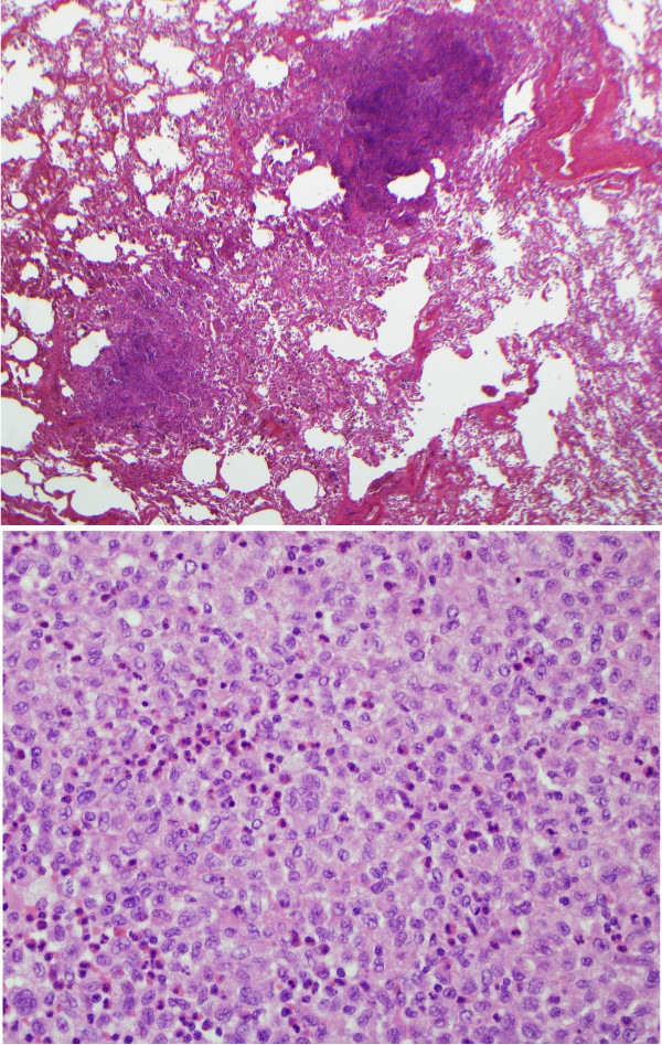
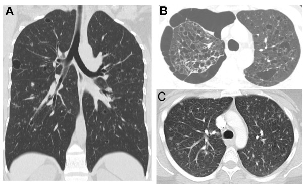
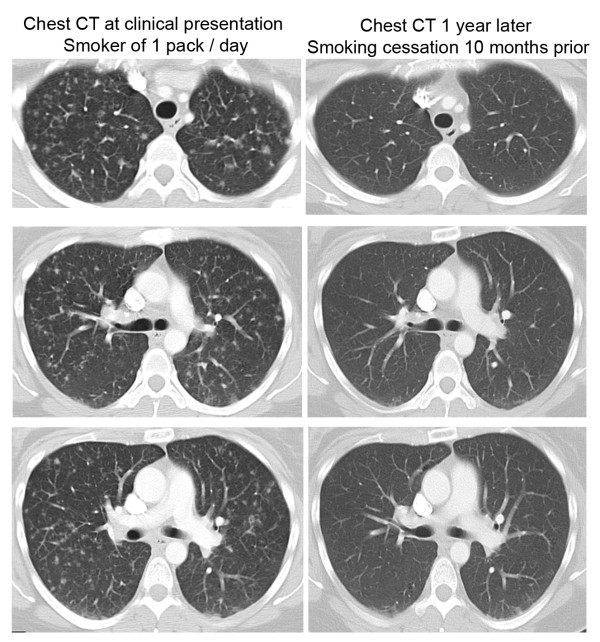
Pulmonary Langerhans Cell Histiocytosis (PLCH) is a relatively uncommon lung disease that generally, but not invariably, occurs in cigarette smokers. The pathologic hallmark of PLCH is the accumulation of Langerhans and other inflammatory cells in small airways, resulting in the formation of nodular inflammatory lesions. While the overwhelming majority of patients are smokers, mechanisms by which smoking induces this disease are not known, but likely involve a combination of events resulting in enhanced recruitment and activation of Langerhans cells in small airways. Bronchiolar inflammation may be accompanied by variable lung interstitial and vascular involvement. While cellular inflammation is prominent in early disease, more advanced stages are characterized by cystic lung destruction, cicatricial scarring of airways, and pulmonary vascular remodeling. Pulmonary function is frequently abnormal at presentation. Imaging of the chest with high resolution chest CT scanning may show characteristic nodular and cystic abnormalities. Lung biopsy is necessary for a definitive diagnosis, although may not be required in instances were imaging findings are highly characteristic. There is no general consensus regarding the role of immunosuppressive therapy in smokers with PLCH. All smokers must be counseled on the importance of smoking cessation, which may result in regression of disease and obviate the need for systemic immunosuppressive therapy. The prognosis for most patients is relatively good, particularly if longitudinal lung function testing shows stability. Complications like pneumothoraces and secondary pulmonary hypertension may shorten life expectancy. Patients with progressive disease may require lung transplantation.


Figures

Figure 1
Figure 1
Proposed pathogenesis of PLCH. The primary event in the pathogenesis probably involves cigarette smoke-induced recruitment and activation of Langerhans cells to the small airways, a process that may result from a variety of potential mechanisms. Cigarette smoke activates epithelial cells and macrophages to produce cytokines and chemokines like granulocyte-macrophage colony-stimulating factor (GM-CSF), Chemokine (C-C motif) ligand 20 (CCL20 or Macrophage Inflammatory Protein-3 alpha), transforming growth factor-β (TGF-β), tumor necrosis factor-α (TNF-α) and osteopontin that promote recruitment, retention and activation of Langerhans cells. Cigarette smoke may also directly activate Langerhans cells. Langerhans cells conditioned by cigarette smoke may inappropriately recognize auto-antigens in the lungs and activate adaptive T cell responses that secondarily mediate injury in airway tissues. Chronic inflammation and cytokine production (particularly TGF-β) may promote local fibroblast activation and airway-centered fibrosis. The combination of airway centered inflammation and tissue remodeling promote dilatation of structures distal to the inflamed small airways and cystic formation. It is possible, although not proven, that autoimmunity directed to antigens expressed by epithelial or other lung cells, may promote premature emphysema. Host genetic factors are likely to be very important in disease development, while the potential role of infection or other environmental factors could be relevant in the induction of disease in some instances (although never proven).
Figure 2
Figure 2
Light microscopy findings in PLCH. Upper panel shows a low-power microscopic picture with nodular airway-centered lesions showing microcystic change (20×, original magnification, hematoxylin and eosin stain). The lower panel shows diffuse infiltration of lung tissue with Langerhans cells showing vesicular nuclear chromatin, irregular nuclear contour and moderate amount of pale cytoplasm devoid of phagocytosed material. Many eosinophils are also intermixed among Langerhans cells (200× magnification, hematoxylin and eosin stain).
Figure 3
Figure 3
Chest CT findings in PLCH. A) Coronal chest CT image from a 36-year-old smoker with biopsy-proven PLCH demonstrating a combination of large and small lung cysts and pulmonary nodules, distributed predominantly in upper and mid-lung zones. B) Chest CT section of a 35-year-old smoker with biopsy-proven PLCH showing extensive bilateral upper lobe cystic lung changes and a medium-sized pneumothorax in the right thoracic cavity. C) Chest CT of a 40-year-old smoker with biopsy-proven PLCH showing a combination of diffuse nodular and cystic abnormalities in both lungs.
Figure 4
Figure 4
PET findings in nodular PLCH. The chest CT images on the left upper and lower panels show multiple lung nodules in a smoker with surgical lung biopsy-proven PLCH. The corresponding PET images on the right upper and lower panels show PET characteristics of the multiple pulmonary nodules. The larger pulmonary nodules (arrowhead) demonstrated intense PET uptake, while other nodules (arrow) are PET-negative (Standardized Uptake Value < 2.5).
Figure 5
Figure 5
Radiographic improvement following smoking cessation. The chest CT images on the left side were performed in an active one pack/day 37 year old smoker with biopsy-proven PLCH. The representative chest CT images demonstrate diffuse nodular infiltrates in both upper and lower lung fields. The patient quit smoking 2 months after the first chest CT was performed. The representative chest CT images on the right side were performed one year after the first chest CT was obtained, and show considerable improvement in the nodular infiltrates following smoking cessation. The patient did not receive corticosteroid or other immunosuppressive therapy.

References

    1. Favara BE, Feller AC, Pauli M, Jaffe ES, Weiss LM, Arico M, Bucsky P, Egeler RM, Elinder G, Gadner H. et al.Contemporary classification of histiocytic disorders. The WHO Committee On Histiocytic/Reticulum Cell Proliferations. Reclassification Working Group of the Histiocyte Society. Med Pediatr Oncol. 1997;29(3):157–166. doi: 10.1002/(SICI)1096-911X(199709)29:3<157::AID-MPO1>3.0.CO;2-C. - DOI - PubMed
    1. Nezelof C, Basset F, Rousseau MF. Histiocytosis × histogenetic arguments for a Langerhans cell origin. Biomedicine. 1973;18(5):365–371. - PubMed
    1. Nezelof C, Basset F, Diebold N. [Histiocytosis X: a differentiated histiocytic process] Pathol Biol (Paris) 1975;23(6):499. - PubMed
    1. Komp DM. Historical perspectives of Langerhans cell histiocytosis. Hematol Oncol Clin North Am. 1987;1(1):9–21. - PubMed
    1. Vassallo R, Ryu JH, Schroeder DR, Decker PA, Limper AH. Clinical outcomes of pulmonary Langerhans'-cell histiocytosis in adults. N Engl J Med. 2002;346(7):484–490. doi: 10.1056/NEJMoa012087. - DOI - PubMed

MeSH terms