. 2012 Mar 19:8:16.
doi: 10.1186/1744-8069-8-16.

Progesterone produces antinociceptive and neuroprotective effects in rats with microinjected lysophosphatidic acid in the trigeminal nerve root

Affiliations

Progesterone produces antinociceptive and neuroprotective effects in rats with microinjected lysophosphatidic acid in the trigeminal nerve root

Min Ji Kim et al. Mol Pain. .

Abstract

Background: In our present study, we studied the role of demyelination of the trigeminal nerve root in the development of prolonged nociceptive behavior in the trigeminal territory.

Results: Under anesthesia, the Sprague-Dawley rats were mounted onto a stereotaxic frame and 3 μL of lysophosphatidic acid (LPA, 1 nmol) was injected into the trigeminal nerve root to produce demyelination. This treatment decreased the air-puff thresholds, persisted until postoperative day 130, and then returned to the preoperative levels 160 days after LPA injection. The LPA-treated rats also showed a significant hyper-responsiveness to pin-prick stimulation. We further investigated the antinociceptive and neuroprotective effects of progesterone in rats undergoing demyelination of the trigeminal nerve root. Progesterone (8, 16 mg/kg/day) was administered subcutaneously, beginning on the operative day, for five consecutive days in the LPA-treated rats. Treatment with progesterone produced significant early anti-allodynic effects and delayed prolonged anti-allodynic effects. The expression of protein zero (P0) and peripheral myelin protein 22 (PMP22) were significantly down-regulated in the trigeminal nerve root on postoperative day 5 following LPA injection. This down-regulation of the P0 and PMP22 levels was blocked by progesterone treatment.

Conclusions: These results suggest that progesterone produces antinociceptive effects through neuroprotective action in animals with LPA-induced trigeminal neuropathic pain. Moreover, progesterone has potential utility as a novel therapy for trigeminal neuropathic pain relief at an appropriate managed dose and is therefore a possible future treatment strategy for improving the recovery from injury.


Figures

Figure 1
Figure 1
Prolonged nociceptive behavior following the microinjection of LPA into the trigeminal nerve root of the rat. (A) Time course analysis of changes in the air-puff thresholds. LPA-treated animals showed significant decreased air-puff thresholds on postoperative day 3. LPA-induced mechanical allodynia was maintained until postoperative day 130. (B) Time course analysis of changes in the pin-prick responses. LPA injection produced significant mechanical hyper-responsiveness to pin-prick stimulation compared with the vehicle-treated group. (C) Time course analysis of changes in the thermal nociception. # P < 0.05, vehicle- vs. LPA-treated group. All points between two identical symbols have the same level of significance.
Figure 2
Figure 2
The morphological changes in the trigeminal nerve root following microinjection of LPA. On postoperative day 1 (A, B) and 14 (C, D), luxol fast blue stained light micrographs reveal severe demyelination of axonal portion of the trigeminal nerve root compared with vehicle-injected root, respectively (E, F). LPA-induced demyelination was recovered on postoperative day 160. Scale bar, 50 μm.
Figure 3
Figure 3
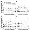
The acute effects of daily treatments with progesterone (8 or 16 mg/kg/day) on mechanical allodynia in rats microinjected with LPA into the trigeminal nerve root. Daily treatment with a low dose of progesterone (8 mg/kg/day) did not produce anti-allodynic effects. However, the administration of 16 mg of progesterone on postoperative days 3, 4, and 5 produced significant anti-allodynic effects. * P < 0.05, vehicle- vs. progesterone-treated group.
Figure 4
Figure 4
Delayed effects of progesterone treatment (8 or 16 mg/kg/day) on mechanical allodynia produced by the microinjection of LPA into the trigeminal nerve root. Progesterone-treated animals showed significant delayed anti-allodynic effects. The lines denote the period of five progesterone treatments. * P < 0.05, vehicle- vs. progesterone-treated group.
Figure 5
Figure 5
The morphological changes in the trigeminal nerve root following treatment of progesterone. Luxol fast blue stained light micrographs reveal the attenuation of demyelination of axonal portion of the trigeminal nerve root compared with vehicle-injected root after daily treatment of progesterone (16 mg/kg/day). Scale bar, 50 μm.
Figure 6
Figure 6
Effects of progesterone treatment (16 mg/kg/day) upon P0 expression. (A) Vehicle treatment did not affect the LPA-induced decreases in P0 expression in the trigeminal nerve root compared with naïve rats on postoperative day 5. However, decreased P0 expression, produced by LPA injection, was recovered following treatment with progesterone. (B) Western blot analysis confirmed that LPA injections cause a decrease in the 27 kDa band for P0, which was recovered by treatment with progesterone. GAPDH was used as a loading control. (C) Quantitative analyses of western blotting data. * P < 0.05, naive- vs. LPA/vehicle-treated group. # P < 0.05, LPA/vehicle-treated group vs. LPA/progesterone-treated group. Scale bar, 50 μm.
Figure 7
Figure 7
Effects of progesterone treatment (16 mg/kg/day) upon PMP22 expression. (A) Vehicle treatments did not affect the LPA-induced decreases in PMP22 expression in the trigeminal nerve root compared with naïve rats on postoperative day 5. However, decreased PMP22 expression, produced by LPA injection, was recovered following treatment with progesterone. (B) Western blot analysis also showed that LPA suppresses the 19 kDa band for PMP22, which was recovered by treatment with progesterone. GAPDH was used as a loading control. (C) Quantitative analyses of western blotting data. * P < 0.05, naive- vs. LPA/vehicle-treated group. # P < 0.05, LPA/vehicle-treated group vs. LAP/progesterone-treated group. Scale bar, 50 μm.

References

    1. Alkayed NJ, Murphy SJ, Traystman RJ, Hurn PD, Miller VM. Neuroprotective effects of female gonadal steroids in reproductively senescent female rats. Stroke. 2000;31:161–168. doi: 10.1161/01.STR.31.1.161. - DOI - PubMed
    1. Roof RL, Duvdevani R, Braswell L, Stein DG. Progesterone facilitates cognitive recovery and reduces secondary neuronal loss caused by cortical contusion injury in male rats. Exp Neurol. 1994;129:64–69. doi: 10.1006/exnr.1994.1147. - DOI - PubMed
    1. Roof RL, Duvdevani R, Heyburn JW, Stein DG. Progesterone rapidly decreases brain edema: treatment delayed up to 24 hours is still effective. Exp Neurol. 1996;138:246–251. doi: 10.1006/exnr.1996.0063. - DOI - PubMed
    1. Shear DA, Galani R, Hoffman SW, Stein DG. Progesterone protects against necrotic damage and behavioral abnormalities caused by traumatic brain injury. Exp Neurol. 2002;178:59–67. doi: 10.1006/exnr.2002.8020. - DOI - PubMed
    1. Azcoitia I, Leonelli E, Magnaghi V, Veiga S, Garcia-Segura LM, Melcangi RC. Progesterone and its derivatives dihydroprogesterone and tetrahydroprogesterone reduce myelin fiber morphological abnormalities and myelin fiber loss in the sciatic nerve of aged rats. Neurobiol Aging. 2003;24:853–860. doi: 10.1016/S0197-4580(02)00234-8. - DOI - PubMed

Publication types

LinkOut - more resources