

Promotion of a functional B cell germinal center response after Leishmania species co-infection is associated with lesion resolution
- PMID: 22429963
- PMCID: PMC3349825
- DOI: 10.1016/j.ajpath.2012.01.012
Promotion of a functional B cell germinal center response after Leishmania species co-infection is associated with lesion resolution
Abstract
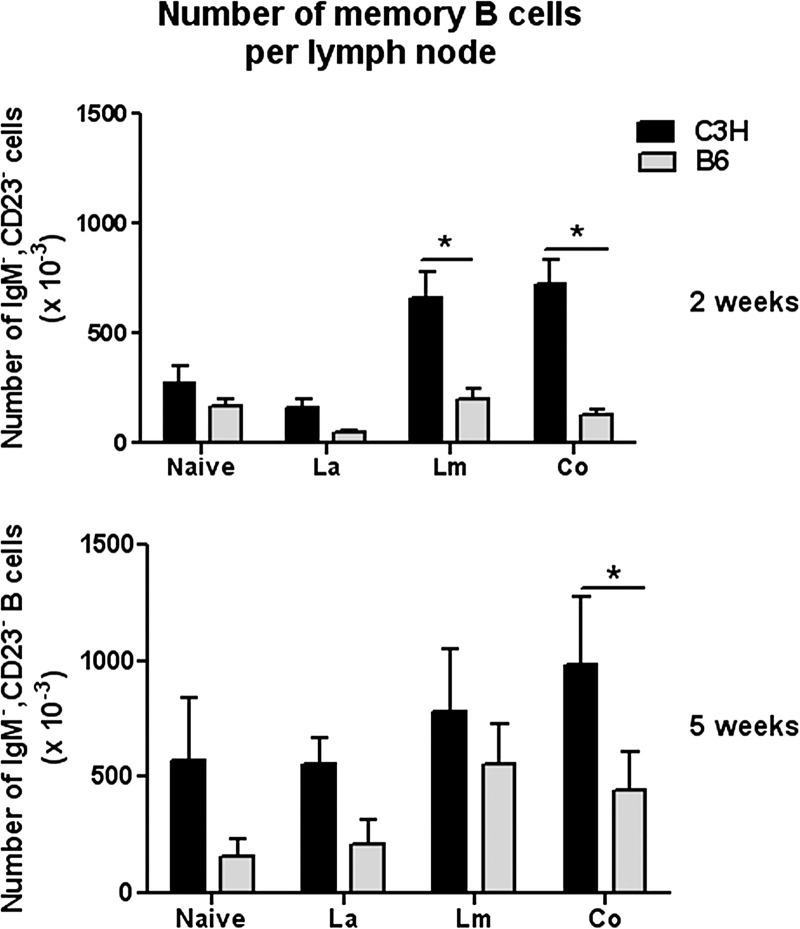
Co-infection of C3HeB/FeJ (C3H) mice with both Leishmania major and Leishmania amazonensis leads to a healed footpad lesion, whereas co-infection of C57BL/6 (B6) mice leads to non-healing lesions. This inability to heal corresponds to a deficiency in B cell stimulation of the macrophage-mediated killing of L. amazonensis in vitro and a less robust antibody response. The mechanism that leads to healing of these lesions is not completely known, although our studies implicate the B cell response as having an important effector function in killing L. amazonensis. To understand more completely this disparate clinical outcome to the same infection, we analyzed the draining lymph node germinal center B cell response between co-infected C3H and B6 mice. There were more germinal center B cells, more antibody isotype-switched germinal center B cells, more memory B cells, and more antigen-specific antibody-producing cells in co-infected C3H mice compared to B6 mice as early as 2 weeks postinfection. Interleukin (IL)-21 production and IL-21 receptor expression in both mouse strains, however, were similar at 2 weeks, suggesting that the difference in the anti-Leishmania response in these mouse strains may be due to differences in T follicular cell commitment or intrinsic B cell differences. These data support the idea that functional B cells are important for healing L. amazonensis in this infectious disease model.
Copyright © 2012 American Society for Investigative Pathology. Published by Elsevier Inc. All rights reserved.
Figures


Similar articles
-
Characterization of the B cell response to Leishmania infection after anti-CD20 B cell depletion.Int J Clin Exp Pathol. 2015 Jun 1;8(6):6192-202. eCollection 2015. Int J Clin Exp Pathol. 2015. PMID: 26261496 Free PMC article.
-
A deficiency in the B cell response of C57BL/6 mice correlates with loss of macrophage-mediated killing of Leishmania amazonensis.Int J Parasitol. 2010 Feb;40(2):157-61. doi: 10.1016/j.ijpara.2009.11.010. Epub 2009 Dec 11. Int J Parasitol. 2010. PMID: 20004204 Free PMC article.
-
Protection of C3HeB/FeJ mice against Leishmania amazonensis challenge after previous Leishmania major infection.Am J Trop Med Hyg. 2004 Oct;71(4):407-11. Am J Trop Med Hyg. 2004. PMID: 15516635
-
Unravelling the contribution of lymph node fibroblasts to vaccine responses.Adv Immunol. 2024;164:1-37. doi: 10.1016/bs.ai.2024.07.001. Epub 2024 Aug 12. Adv Immunol. 2024. PMID: 39523027 Review.
-
Deciphering the Human Germinal Center: A Review of Models to Study T-B Cell Interactions.Eur J Immunol. 2025 Feb;55(2):e202451460. doi: 10.1002/eji.202451460. Eur J Immunol. 2025. PMID: 39931794 Free PMC article. Review.
Cited by
-
Tertiary lymphoid structures in pulmonary granulomas of cattle experimentally infected with aerosolized Mycobacterium bovis.BMC Vet Res. 2025 Jun 5;21(1):403. doi: 10.1186/s12917-025-04804-x. BMC Vet Res. 2025. PMID: 40468399 Free PMC article.
-
CD4 T cell activation by B cells in human Leishmania (Viannia) infection.BMC Infect Dis. 2014 Feb 25;14:108. doi: 10.1186/1471-2334-14-108. BMC Infect Dis. 2014. PMID: 24568275 Free PMC article.
-
Characterization of the B cell response to Leishmania infection after anti-CD20 B cell depletion.Int J Clin Exp Pathol. 2015 Jun 1;8(6):6192-202. eCollection 2015. Int J Clin Exp Pathol. 2015. PMID: 26261496 Free PMC article.
-
Host and parasite responses in human diffuse cutaneous leishmaniasis caused by L. amazonensis.PLoS Negl Trop Dis. 2019 Mar 7;13(3):e0007152. doi: 10.1371/journal.pntd.0007152. eCollection 2019 Mar. PLoS Negl Trop Dis. 2019. PMID: 30845223 Free PMC article.
-
The role of B cells and humoral immunity in Mycobacterium tuberculosis infection.Semin Immunol. 2014 Dec;26(6):588-600. doi: 10.1016/j.smim.2014.10.005. Epub 2014 Oct 28. Semin Immunol. 2014. PMID: 25458990 Free PMC article. Review.
References
-
- Vanloubbeeck Y., Jones D.E. Protection of C3HeB/FeJ mice against Leishmania amazonensis challenge after previous Leishmania major infection. Am J Trop Med Hyg. 2004;71:407–411. - PubMed
-
- Veras P., Brodskyn C., Balestieri F., Freitas L., Ramos A., Queiroz A., Barral A., Beverley S., Barral-Netto M. A dhfr-ts- Leishmania major knockout mutant cross-protects against Leishmania amazonensis. Mem Inst Oswaldo Cruz. 1999;94:491–496. - PubMed
-
- Mukbel R., Petersen C.A., Jones D.E. Soluble factors from Leishmania major-specific CD4(+)T cells and B cells limit L. amazonensis amastigote survival within infected macrophages. Microbes Infect. 2006;8:2547–2555. - PubMed
Publication types
MeSH terms
Substances
Grants and funding
LinkOut - more resources
Full Text Sources
