Case Reports
. 2012 Jul;27(7):1462-70.
doi: 10.1002/jbmr.1603.

Denosumab treatment for fibrous dysplasia

Affiliations
Case Reports

Denosumab treatment for fibrous dysplasia

Alison M Boyce et al. J Bone Miner Res. 2012 Jul.

Abstract

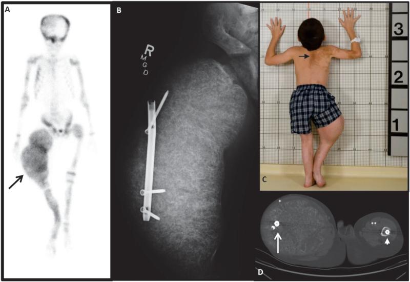
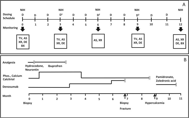
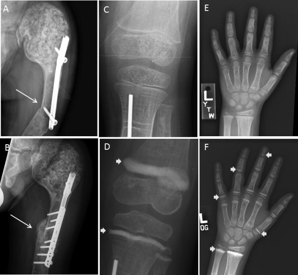
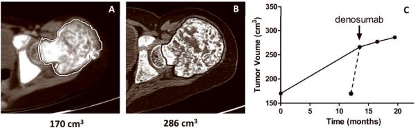
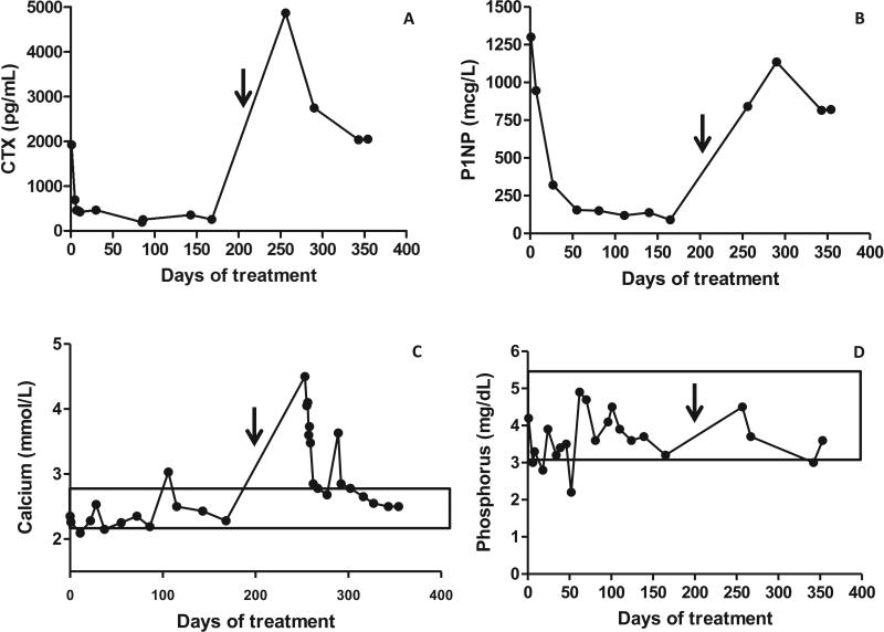
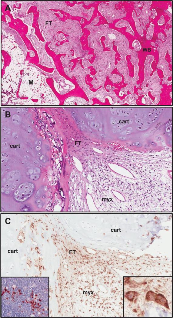
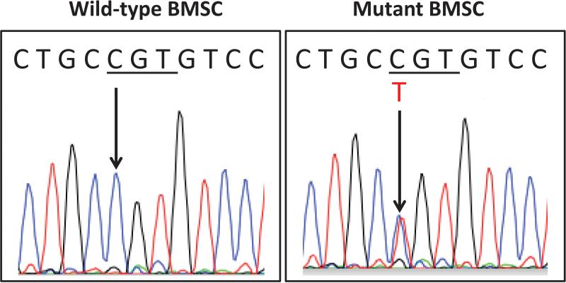
Fibrous dysplasia (FD) is a skeletal disease caused by somatic activating mutations of the cyclic adenosine monophosphate (cAMP)-regulating protein, α-subunit of the Gs stimulatory protein (G(s) α). These mutations lead to replacement of normal bone by proliferative osteogenic precursors, resulting in deformity, fracture, and pain. Medical treatment has been ineffective in altering the disease course. Receptor activator of NF-κB ligand (RANKL) is a cell-surface protein involved in many cellular processes, including osteoclastogenesis, and is reported to be overexpressed in FD-like bone cells. Denosumab is a humanized monoclonal antibody to RANKL approved for treatment of osteoporosis and prevention of skeletal-related events from bone metastases. We present the case of a 9-year-old boy with severe FD who was treated with denosumab for a rapidly expanding femoral lesion. Immunohistochemical staining on a pretreatment bone biopsy specimen revealed marked RANKL expression. He was started on monthly denosumab, with an initial starting dose of 1 mg/kg and planned 0.25 mg/kg dose escalations every 3 months. Over 7 months of treatment he showed marked reduction in pain, bone turnover markers (BTMs), and tumor growth rate. Denosumab did not appear to impair healing of a femoral fracture that occurred while on treatment. With initiation of treatment he developed hypophosphatemia and secondary hyperparathyroidism, necessitating supplementation with phosphorus, calcium, and calcitriol. BTMs showed rapid and sustained suppression. With discontinuation there was rapid and dramatic rebound of BTMs with cross-linked C-telopeptide (reflecting osteoclast activity) exceeding pretreatment levels, accompanied by severe hypercalcemia. In this child, denosumab lead to dramatic reduction of FD expansion and FD-related bone pain. Denosumab was associated with clinically significant disturbances of mineral metabolism both while on treatment and after discontinuation. Denosumab treatment of FD warrants further study to confirm efficacy and determine potential morbidity, as well as to determine the mechanism of RANKL in the pathogenesis of FD and related bone marrow stromal cell diseases.


Figures

Figure 1
Figure 1. Initial Clinical Presentation
A) 99Technectium-MDP bone scan showing extensive tracer uptake at multiple areas of FD and ballooning expansion of the right femur (arrow). B) X-ray shows massive expansion of right femur and displacement of a rod that was originally in the intramedullary canal. C) Photograph showing classic “coast-of-Maine” café-au-lait spots (arrow) and the effect of massive overgrowth of the right femur. D) CT at the level of the femura mid-shafts, with lateral displacement of the rod on the right (white arrow), and an intramedullary rod on the left (arrow head). The quadriceps muscles on the right have been stretched to dysfunctional bands (*), compared to the left (**).
Figure 2
Figure 2. Proposed treatment regimen and clinical course
A) Proposed Regimen. Dosing regimen and monitoring schedule are indicated. Denosumab was to be given monthly at an initial dose of 1 mg/kg. Dose escalations were planned at three month intervals (blue arrows) to 1.25 mg/kg, 1.5 mg/kg, and 1.75 mg/kg. The patient was evaluated by his local endocrinologist monthly and at NIH every three months. Mineral panels were monitored weekly for the first three months and then monthly (not shown). Bone turnover markers were monitored monthly (not shown). The scale represents months. D = dose of denosumab, TV = tumor volume, AS = arm spam, XR = Hand XR, DE = dental exam, BX = bone biopsy. B) Clinical Course. The periods for the administration of analgesics, supplemental phosphorus, calcium and calcitriol, denosumab, and bisphosphonate treatment (pamidronate or zoledronic acid) are indicated by the transverse lines, and cessation by the double vertical lines. Time points for biopsy, fracture and start of hypercalcemia are indicated. The scale represents months.
Figure 3
Figure 3. Radiographs
A) A radiograph at the time of fracture indicated the fracture occurred at the distal end of a previously intramedullary rod (longer arrow). B. Eight weeks after lplate fixation shows callus formation at the fracture site (arrow). C&D) Knee radiographs pre-treatment and 6 months after the initiation of treatment, as indicated. E&F) Hand radiographs, as above. Note the thick sclerotic bands at the level of the metaphyses (arrows), reminiscent of the radiologic appearance of children treated with bisphosphonates. There does not appear to be evidence of frank rickets.
Figure 4
Figure 4. Tumor volume
A) A representative two dimensional image created from a horizontal slice prior to denosumab treatment at the level of the femoral head taken from a thin slice CT study that was used to measure tumor volume (see Methods). B) A similar image taken two months after the final dose of denosumab. C) This panel shows the change in tumor volume over time. The first and last time points were derived from the studies done for panels A&B. The start of denosumab treatment is indicated. There were 13.5 months between the initial study and the scan done prior to start of denosumab. The solid line represents the change in volume assuming a linear rate of growth. Per report of the patient's mother and orthopedic surgeon, expansion began acutely and progressed rapidly over six weeks prior to initiation of denosumab, represented by the dashed line. Initiation of treatment was associated with marked decrease in the rate of tumor growth.
Figure 5
Figure 5. Biochemical Response to Treatment
A) Serum B-CTX, a marker of bone resorption. B) Serum P1NP, a marker of bone formation. C) Serum calcium. D) Serum phosphorus. Denosumab was initiated at Day 0 and discontinued at day 210, indicated by the arrow. After discontinuation of denosumab there was a marked increase in BTMs, especially the resorption marker CTX that was associated with marked hypercalcemia in the presence of a suppressed serum PTH, and 1,25 (OH)2 vitamin D. Normal ranges for calcium and phosphorus are indicated by boxes. Normal ranges for CTX and P1NP in children are not well defined.
Figure 6
Figure 6. Histopathology
A) An H&E stained section of FD taken prior to the initiation of denosumab from a region of the lesion that demonstrated typical features of FD. The overall “Chinese character” appearance is evident and consists of areas of typical fibrous tissue (FT) interspersed amongst trabecular structures composed of woven bone (WB), as well as small region of relatively fatty bone marrow (M). B) This H&E section was from a region of the lesion that had a prominent cartilaginous component (cart), as well as areas of FT composed of stromal cells and an area with a myxoid appearance (myx), which again is sometimes seen in FD, but is not typical. C) A serial section adjacent to the area shown in panel B stained for RANKL demonstrated that most of the stromal cells were positive for RANKL with a cell surface staining pattern, and that the chondroid cells were negative. The inset panel on the right shows a high power view of RANKL positive stromal cells and the inset panel on the left from a normal human lymph node with RANKL positive lymphoid cells served as a positive control for RANKL staining.
Figure 7
Figure 7. Mutation Analysis
BMSCs were isolated from a pre-treatment bone biopsy and expanded in vitro, as described in the Methods section. Total DNA was isolated and the mutated Gsα-specific region was amplified. PCR products were sequenced, and DNA sequencing from the Arginine-201 region is shown. Clonal strains carrying the wild-type Gsα (C G T) or the heterozygous R201C mutation (C/T G T) are indicated by arrows.

References

    1. Collins MT, Bianco P. Fibrous dysplasia. In: Favus MJ, editor. Primer on the Metabolic Bone Diseases and Disorders of Mineral Metabolism. 6ed. American Society for Bone and Mineral Research; Washington, D.C.: 2006. pp. 415–418.
    1. Lichtenstein L. Poloyostotic fibrous dysplasia. Arch Surg. 1938;36:874–898.
    1. Lichtenstein L, Jaffe HL. Fibrous dysplasia of bone: a condition affecting one, several or many bones, the graver cases of which may present abnormal pigmentation of skin, premature sexual development, hyperthyroidism or still other extraskeletal abnormalities. ArchPathol. 1942;33:777–816.
    1. McCune DJ. Osteitis fibrosa cystica; the case of a nine year old girl who also exhibits precocious puberty, multiple pigmentation of the skin and hyperthyroidism. Am J Dis Child. 1936;52:743–744.
    1. Albright F, Butler AM, Hampton AO, Smith PH. Syndrome characterized by osteitis fibrosa disseminata, areas of pigmentation and endocrine dysfunction, with precocious puberty in females, report of five cases. N Engl J Med. 1937;216:727–746.

Publication types