. 2012 Jun 14;119(24):5706-14.
doi: 10.1182/blood-2011-12-397489. Epub 2012 Mar 19.

Autonomous murine T-cell progenitor production in the extra-embryonic yolk sac before HSC emergence

Affiliations

Autonomous murine T-cell progenitor production in the extra-embryonic yolk sac before HSC emergence

Momoko Yoshimoto et al. Blood. .

Abstract

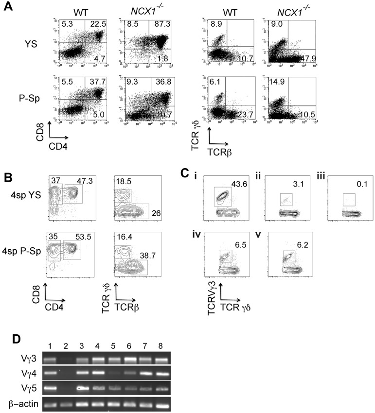
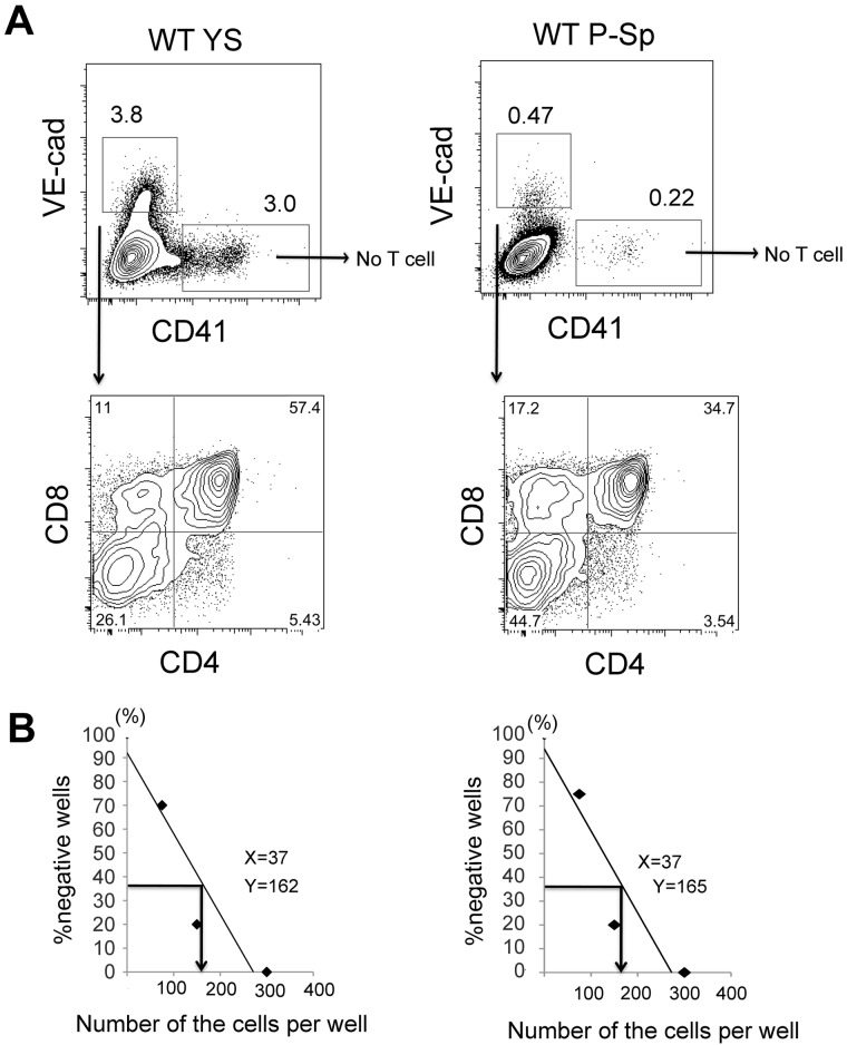
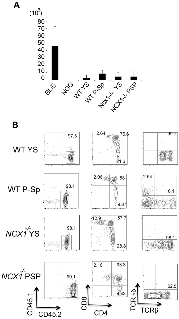
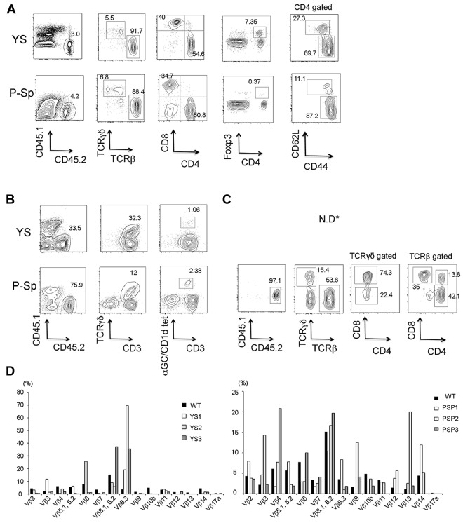
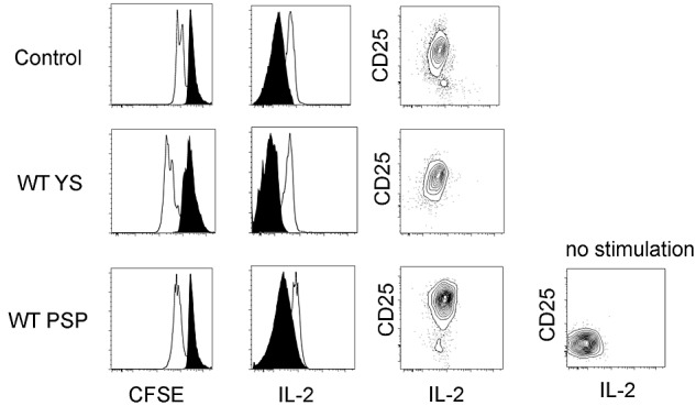
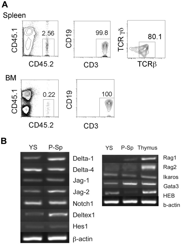
The extra-embryonic yolk sac (YS) is the first hematopoietic site in the mouse embryo and is thought to generate only primitive erythroid and myeloerythroid progenitor cells before definitive HSC emergence within the embryo on E10.5. Here, we have shown the existence of T cell-restricted progenitors in the E9.5 YS that directly engraft in recipient immunodeficient mice. T-cell progenitors were also produced in vitro from both YS and para-aortic splanchnopleura hemogenic endothelial cells, and these T-cell progenitors repopulated the thymus and differentiated into mature T-cell subsets in vivo on transplantation. Our data confirm that the YS produces T-lineage-restricted progenitors that are available to colonize the thymus and provide new insight into the YS as a definitive hematopoietic site in the mouse embryo.


Figures

Figure 1
Figure 1
YS and P-Sp have autonomous T-cell potential. (A) E9.5 YS and P-Sp cells from WT and Ncx1−/− embryos were plated on OP9-DL1 stromal cells with added IL-7. After 10-14 days of coculture, CD4+CD8+ DP cells as well as TCRβ and TCRγδ+ were produced in each culture. (B) YS and P-Sp cells at 4-sp stage also produced DP as well as CD8+ cells in OP9-DL1 coculture. Representative FACS dot plots are depicted for n = 5 in each YS and P-Sp grouping. (C) TCRVγ3 expression was examined by flow cytometry with (i) E15.5 fetal thymocytes, (ii) cells derived from E14.5 fetal liver linc-kit+Sca-1+ (KSL) cells in OP9-DL1 culture, (iii) cells derived from adult BM KSL cells in OP9-DL1 culture, (iv) E9.5 YS-derived cells, and (v) E9.5 P-Sp–derived cells. TCRγδ+ cells were gated for each dot plot analysis. (D) RT-PCR analysis detecting TCR Vγ3, Vγ4, and Vγ5 transcripts. (1) Day 10 neonatal thymocytes, (2) E15.5 thymocytes, and cultured cells in OP9-DL1 derived from (3) 4 sp YS, (4) 4 sp P-Sp, (5) E9.5 YS, (6) E9.5 P-Sp, (7) E14.5 fetal liver KSL, and (8) E15.5 fetal liver common lymphoid progenitor (CLP) cells.
Figure 2
Figure 2
VE-cad+CD41 hemogenic endothelial cells produce T cells in both YS and P-Sp with similar frequency. (A) VE-cad+CD41 or VE-cadCD41+ cells were sorted from E9.5 WT YS and P-Sp and were plated on OP9-DL1 stromal cells in a limited dilution manner (75-300 cells/well). After 7-10 days, each well containing cobblestone-forming area and lymphocyte-like cells was analyzed by flow cytometry. (B) CD4+CD8+DP cells were obtained from VE-cad+CD41 cell population, not from VE-cadCD41+ cells at a frequency of 1 of 162 YS VE-cad+ cells (left panel) and 1 of 165 P-Sp VE-cad+ cells (right panel), respectively.
Figure 3
Figure 3
Recipient thymus was transiently reconstituted by YS/P-Sp–derived progenitor cells. (A) YS and P-Sp cells from Ncx1−/− and WT embryos were cocultured with OP9-DL1 for 10-12 days and transplanted into the peritoneal cavity of sublethally irradiated NOG neonates. Two weeks after injection, recipient thymuses were analyzed. Although there was no apparent structural thymus in NOG neonates, mice that received a transplant display an identifiable thymus that contains ∼ 1 × 10⁶ cells. When analyzed at 14 weeks after injection, no thymus was detected in any mice that received a transplant (Table 1). The cell numbers of the thymus from C57BL/6 mice that did not receive a transplant were enumerated as a control. (B) FACS analysis of reconstituted thymus from recipient mice. All the thymocytes were donor derived and consisted of CD4+CD8+ DP cells as well as CD4+ SP and CD8+ SP cells, with TCRβ expression. The number of mice that received a transplant is shown in Table 1.
Figure 4
Figure 4
YS- and P-Sp–derived DN T cells can differentiate into distinct T-cell subsets in vivo. Cells from the recipient spleen (A) and liver (B) were analyzed for the presence of mature T-cell populations 2 weeks after transplantation. Memory (CD62LlowCD44high) T cells were analyzed 14 weeks after injection (A right panels, dot plot is CD45.2+CD4+ gated). Regulatory (CD4+FoxP3+) T cells, naive (CD4+CD62LhighCD44low) T cells, and memory (CD4+CD62LlowCD44high) T cells were detected in the recipient spleen (A), and γδT (TCRγδ+CD3+), and α-Gal–loaded CD1d tetramer+ (αGC/CD1d tet+) CD3+ NKT cells were detected in the recipient liver (B). (C) TCRβ+ or TCRγδ+CD8+ intraepithelial T cells were also detected in the intestine of a mouse that received a transplant with P-Sp–derived T cells. ND indicates not determined for YS-derived cells. The representative FACS dot plots are depicted. The number of mice that received a transplant is detailed in Table 1. (D) The TCRVβ repertoires of YS-derived (left) and P-Sp–derived (right) CD3+ T cells engrafted in the recipient spleen are depicted (each n = 3).
Figure 5
Figure 5
YS- and P-Sp–derived T cells engrafted in the recipient spleen are functional. YS- and P-Sp–derived T cells engrafted in the recipient spleen are proliferative on CD3 stimulation and secrete IL-2. Spleen cells from recipient mice 2 weeks after transplantation were plated in RPMI medium with anti-CD3 and anti-CD28 Abs. Donor cell proliferation was analyzed with the use of CFSE staining (left panel: CD45.2+CD4+ gated cells are depicted; CD45.2+CD8+ cells showed a similar proliferation pattern). Nontransplanted BL/6 spleen cells were used as a control. IL-2 secretion was confirmed by intracellular staining (middle panel). CD25 expression was up-regulated in all samples (right panel). Nonstimulated spleen cells were used as a negative control. Filled histogram is at 0 hour, and solid line is at 48 hours after stimulation. YS- and P-Sp–derived spleen cells (n = 5).
Figure 6
Figure 6
YS contains unipotent T-cell progenitors that can engraft in the NOG neonates. (A) E9.5 YS reconstituted only T cells in the recipient spleen and BM on transplantation. Most of CD3+ cells were TCRβ+. The representative FACS dot plots from a recipient mouse 9 months after transplantation are depicted. The number of mice that received a transplant is shown in supplemental Table 3. (B) RT-PCR was performed with E9.5 WT YS and P-Sp for Notch ligand. The expression of Notch signaling genes (B left panel) and of genes that are important for T lymphoid development (B right panel) are shown.

Comment in

  • The puzzling origin of lymphocytes.
    Muller-Sieburg C. Muller-Sieburg C. Blood. 2012 Jun 14;119(24):5609-10. doi: 10.1182/blood-2012-04-420737. Blood. 2012. PMID: 22700689 No abstract available.

References

    1. Keller G. Embryonic stem cell differentiation: emergence of a new era in biology and medicine. Genes Dev. 2005;19(10):1129–1155. - PubMed
    1. Moore MA, Metcalf D. Ontogeny of the haemopoietic system: yolk sac origin of in vivo and in vitro colony forming cells in the developing mouse embryo. Br J Haematol. 1970;18(3):279–296. - PubMed
    1. Lux CT, Yoshimoto M, McGrath K, Conway SJ, Palis J, Yoder MC. All primitive and definitive hematopoietic progenitor cells emerging before E10 in the mouse embryo are products of the yolk sac. Blood. 2008;111(7):3435–3438. - PMC - PubMed
    1. Tober J, McGrath KE, Palis J. Primitive erythropoiesis and megakaryopoiesis in the yolk sac are independent of c-myb. Blood. 2008;111(5):2636–2639. - PMC - PubMed
    1. Muller AM, Medvinsky A, Strouboulis J, Grosveld F, Dzierzak E. Development of hematopoietic stem cell activity in the mouse embryo. Immunity. 1994;1(4):291–301. - PubMed

Publication types

MeSH terms