

Whole-brain, time-locked activation with simple tasks revealed using massive averaging and model-free analysis
- PMID: 22431587
- PMCID: PMC3325687
- DOI: 10.1073/pnas.1121049109
Whole-brain, time-locked activation with simple tasks revealed using massive averaging and model-free analysis
Abstract
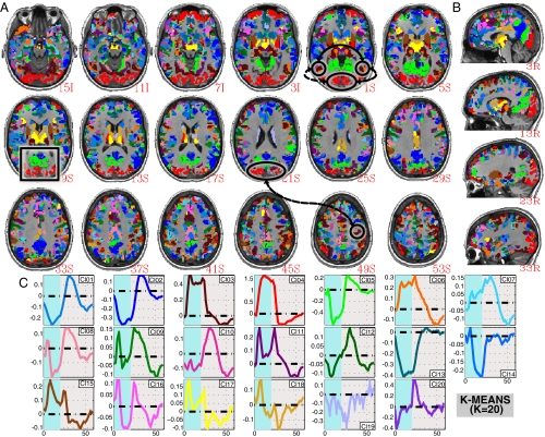
The brain is the body's largest energy consumer, even in the absence of demanding tasks. Electrophysiologists report on-going neuronal firing during stimulation or task in regions beyond those of primary relationship to the perturbation. Although the biological origin of consciousness remains elusive, it is argued that it emerges from complex, continuous whole-brain neuronal collaboration. Despite converging evidence suggesting the whole brain is continuously working and adapting to anticipate and actuate in response to the environment, over the last 20 y, task-based functional MRI (fMRI) have emphasized a localizationist view of brain function, with fMRI showing only a handful of activated regions in response to task/stimulation. Here, we challenge that view with evidence that under optimal noise conditions, fMRI activations extend well beyond areas of primary relationship to the task; and blood-oxygen level-dependent signal changes correlated with task-timing appear in over 95% of the brain for a simple visual stimulation plus attention control task. Moreover, we show that response shape varies substantially across regions, and that whole-brain parcellations based on those differences produce distributed clusters that are anatomically and functionally meaningful, symmetrical across hemispheres, and reproducible across subjects. These findings highlight the exquisite detail lying in fMRI signals beyond what is normally examined, and emphasize both the pervasiveness of false negatives, and how the sparseness of fMRI maps is not a result of localized brain function, but a consequence of high noise and overly strict predictive response models.
Conflict of interest statement
The authors declare no conflict of interest.
Figures


References
-
- Biswal B, Yetkin FZ, Haughton VM, Hyde JS. Functional connectivity in the motor cortex of resting human brain using echo-planar MRI. Magn Reson Med. 1995;34:537–541. - PubMed
-
- Cherkassky VL, Kana RK, Keller TA, Just MA. Functional connectivity in a baseline resting-state network in autism. Neuroreport. 2006;17:1687–1690. - PubMed
MeSH terms
LinkOut - more resources
Full Text Sources
Other Literature Sources
