. 2012 Apr 3;109(14):5223-8.
doi: 10.1073/pnas.1118699109. Epub 2012 Mar 19.

Structure of a hepatitis C virus RNA domain in complex with a translation inhibitor reveals a binding mode reminiscent of riboswitches

Affiliations

Structure of a hepatitis C virus RNA domain in complex with a translation inhibitor reveals a binding mode reminiscent of riboswitches

Sergey M Dibrov et al. Proc Natl Acad Sci U S A. .

Abstract

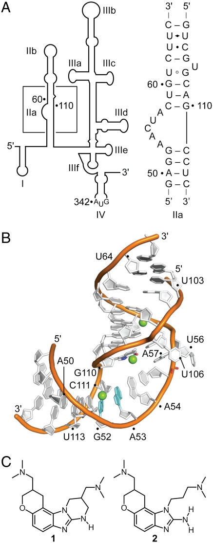
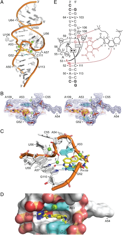
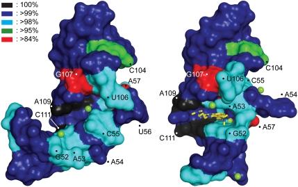
The internal ribosome entry site (IRES) in the hepatitis C virus (HCV) RNA genome is essential for the initiation of viral protein synthesis. IRES domains adopt well-defined folds that are potential targets for antiviral translation inhibitors. We have determined the three-dimensional structure of the IRES subdomain IIa in complex with a benzimidazole translation inhibitor at 2.2 Å resolution. Comparison to the structure of the unbound RNA in conjunction with studies of inhibitor binding to the target in solution demonstrate that the RNA undergoes a dramatic ligand-induced conformational adaptation to form a deep pocket that resembles the substrate binding sites in riboswitches. The presence of a well-defined ligand-binding pocket within the highly conserved IRES subdomain IIa holds promise for the development of unique anti-HCV drugs with a high barrier to resistance.


Conflict of interest statement

The authors declare no conflict of interest.

Figures

Fig. 1.
Fig. 1.
The HCV IRES RNA target. (A) Secondary structure of the 5′ UTR (nucleotides 1–341 of HCV genotype 1b). The boxed region indicates the subdomain IIa whose sequence is shown. (B) Crystal structure of the subdomain IIa (10). Mg2+ ions are shown as green spheres. (C) Benzimidazole translation inhibitors of the HCV IRES (15, 17).
Fig. 2.
Fig. 2.
Crystal structure of the subdomain IIa RNA inhibitor complex. (A) Overall view of the complex. The benzimidazole ligand (2) is in yellow. Mg2+ ions are shown as green spheres. (B) Stereoview of the 2Fo-Fc electron density contoured at 1σ around the ligand-binding site. (C) Detail view of the ligand-binding site. The bases of G52 and A53, which form the intercalation site for the benzimidazole scaffold, are shown in cyan. The purine of G110 provides the docking edge for the amino-imidazole group. Hydrogen bond interactions are indicated by dashed lines. (D) Surface representation, highlighting the deep ligand-binding pocket. (E) Schematic of the interactions in the ligand-binding site (SI Appendix, Table S3). Hydrogen bonds are shown as dashed lines. Formation of non-Watson–Crick base pairs is indicated with solid lines and symbols according to Leontis et al. (45). Stacked lines (≡) indicate stacking of bases and intercalation of the ligand. Residues interacting with the benzimidazole are highlighted in red.
Fig. 3.
Fig. 3.
Binding site conservation in the HCV IRES target. The sequence conservation in clinical isolates of HCV has been mapped on the surface of the RNA structure. (Left) The unliganded RNA, (Right) the complex with the viral translation inhibitor 2. The degree of conservation is indicated by color coding. See also SI Appendix, Table S3, Fig. S6).
Fig. 4.
Fig. 4.
Binding and biological activity of benzimidazole 2 at the HCV IRES target. (A) Fluorescence signal for a titration of 2AP54-labeled IIa RNA with compound 2 in the presence of 10 mM Mg2+. Error bars represent ± 1 SD calculated from three independent titrations. Fitting of a dose-response curve resulted in an EC50 value of 12 ± 1 μM. (B) FRET signal for titrations of Cy3/Cy5-labeled IIa RNA with benzimidazole 2 in the presence of 2 mM Mg2+. Symbols represent WT RNA (○), A57U mutant (∇), and two double mutants, C58G/G110C (□) and C58U/G110A (◊). Fitting of dose-response curves resulted in EC50 values for ligand binding of 3.4 ± 0.3 μM (WT) and 9.3 ± 1.1 μM (A57U). The double mutants did not bind the inhibitor (EC50 > 100 μM). (C) FRET signal for a titration of Cy3/Cy5-labeled IIa WT RNA with the analog compound 3 that has the 2-amino functionality replaced by a methyl group. The analog does not bind the RNA target. (D) Inhibition of HCV translation in human Huh-7.5 cells. Titrations of compound 2 were performed against BM4-5 FEO subgenomic replicon (•) and JFH1 full-length virus (○). Fitting of dose-response curves resulted in EC50 values for translation inhibition of 2.8 ± 0.4 μM (replicon) and 3.4 ± 0.5 μM (virus). In all graphs error bars represent ± 1 SD calculated from triplicate experiments, except for D where four physical replicates of triplicate experiments were used.

References

    1. Jang JY, Chung RT. Chronic hepatitis C. Gut Liver. 2011;5:117–132. - PMC - PubMed
    1. Enserink M. Infectious diseases. First specific drugs raise hopes for hepatitis C. Science. 2011;332:159–160. - PubMed
    1. Feld JJ, Hoofnagle JH. Mechanism of action of interferon and ribavirin in treatment of hepatitis C. Nature. 2005;436:967–972. - PubMed
    1. Robinson M, Tian Y, Delaney WEt, Greenstein AE. Preexisting drug-resistance mutations reveal unique barriers to resistance for distinct antivirals. Proc Natl Acad Sci USA. 2011;108:10290–10295. - PMC - PubMed
    1. Davis DR, Seth PP. Therapeutic targeting of HCV internal ribosomal entry site RNA. Antiviral Chem Chemother. 2011;21:117–128. - PubMed

Publication types

Associated data