. 2012 Apr 3;109(14):5247-52.
doi: 10.1073/pnas.1117268109. Epub 2012 Mar 19.

Effect of channel mutations on the uptake and release of the retinal ligand in opsin

Affiliations

Effect of channel mutations on the uptake and release of the retinal ligand in opsin

Ronny Piechnick et al. Proc Natl Acad Sci U S A. .

Abstract

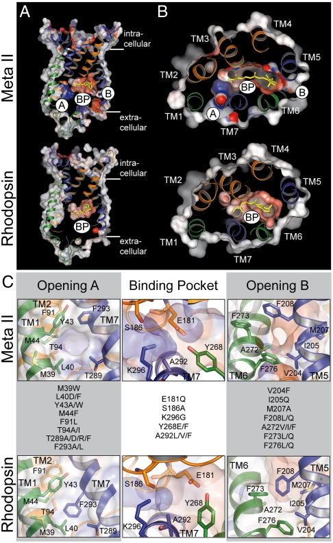
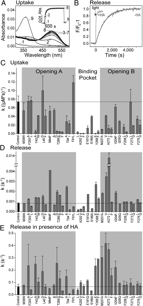
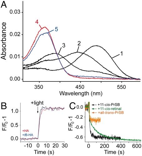
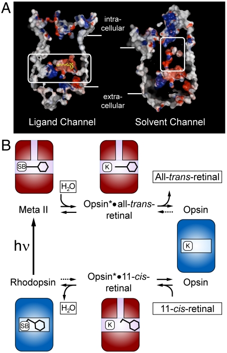
In the retinal binding pocket of rhodopsin, a Schiff base links the retinal ligand covalently to the Lys296 side chain. Light transforms the inverse agonist 11-cis-retinal into the agonist all-trans-retinal, leading to the active Meta II state. Crystal structures of Meta II and the active conformation of the opsin apoprotein revealed two openings of the 7-transmembrane (TM) bundle towards the hydrophobic core of the membrane, one between TM1/TM7 and one between TM5/TM6, respectively. Computational analysis revealed a putative ligand channel connecting the openings and traversing the binding pocket. Identified constrictions within the channel motivated this study of 35 rhodopsin mutants in which single amino acids lining the channel were replaced. 11-cis-retinal uptake and all-trans-retinal release were measured using UV/visible and fluorescence spectroscopy. Most mutations slow or accelerate both uptake and release, often with opposite effects. Mutations closer to the Lys296 active site show larger effects. The nucleophile hydroxylamine accelerates retinal release 80 times but the action profile of the mutants remains very similar. The data show that the mutations do not probe local channel permeability but rather affect global protein dynamics, with the focal point in the ligand pocket. We propose a model for retinal/receptor interaction in which the active receptor conformation sets the open state of the channel for 11-cis-retinal and all-trans-retinal, with positioning of the ligand at the active site as the kinetic bottleneck. Although other G protein-coupled receptors lack the covalent link to the protein, the access of ligands to their binding pocket may follow similar schemes.


Conflict of interest statement

The authors declare no conflict of interest.

Figures

Fig. 1.
Fig. 1.
Comparison of the inactive rhodopsin and active Meta II crystal structures and location of the residues mutated in this study. (A) longitudinal and (B) coplanar (cytoplasmic view) cuts through Meta II (top; PDB accession 3PXO) and rhodopsin (bottom; PDB accession 1U19). In both boxes opening A (A) between TM1 and TM7, the retinal binding pocket (BP), and opening B (B) between TM 5 and TM 6 are assigned. Electrostatic surface potentials are contoured at ± 30 kT/e with negatively and positively charged surface areas in red and blue, respectively. (C) Close-ups of openings A and B (side views) and of the retinal binding pocket (top view) with the amino acid residues mutated in this study as indicated.
Fig. 2.
Fig. 2.
Kinetics of retinal uptake and release in rhodopsin mutants. (A) Regeneration of N2C/D282C opsin (control) with 11-cis-retinal monitored by UV/visible spectroscopy: spectrum of 0.5 μM opsin (1), 30 s after addition of 1 μM 11-cis-retinal (2) and spectra recorded subsequently every 60 s (–8). Spectrum 9 was taken after addition of 25 mM hydroxylamine and illumination of the sample. The inset shows a plot of the absorbance change at 500 nm as a function of time (numbers identify respective spectra). The solid line is a fit to the data points with a bimolecular rate equation (see SI Text: SI Materials and Methods). (B) Fluorescence spectroscopic analysis of light-induced all-trans-retinal release from N2C/D282C rhodopsin (control) in the absence (−HA) and in the presence of 25 mM hydroxylamine (+HA). Reactions were initiated by 15 s illumination with orange light. Reaction rates were determined by a monoexponential fit to the data points (−HA, solid line) or by normalizing the initial slope of the fluorescence change to the maximum amplitude (+HA). (C) Regeneration rates of opsin mutants measured as illustrated in (A). Light-induced all-trans-retinal release rates of rhodopsin mutants measured in the absence (D) or in the presence of HA (E) as illustrated in (B). The dashed lines in (C–E) refer to the values obtained for N2C/D282C rhodopsin (control) and error bars display SD of five (control) or three (all other mutants) independent measurements. All measurements were performed at pH 6.0, 20 °C in 0.03% DDM.
Fig. 3.
Fig. 3.
Characterization of mutant K296G. (A) UV/visible spectrum of K296G opsin regenerated with 11-cis-PrSB in the dark (1), 30 s (2) and 160 min (3) after illumination of the sample. Spectra (4) and (5) were recorded 30 s after illumination of samples containing 25 mM hydroxylamine or tB-HA, respectively. (B) Light-induced retinal release from K296G monitored by fluorescence spectroscopy in the absence (black trace), and in the presence of 25 mM hydroxylamine (red trace) or tB-HA (blue trace), respectively. (C) Fluorescence change induced by addition of 1 μM 11-cis-PrSB (black trace) or all-trans-PrSB (orange trace) to 0.5 μM K296G opsin. Addition of 1 μM 11-cis-retinal to control opsin (green trace). All measurements were performed at pH 6.0, 10 °C in 0.03% DDM.
Fig. 4.
Fig. 4.
Model of opening and closing of the retinal channel and its relation to the activation/deactivation cycle of rhodopsin. (A) Putative retinal (left) and solvent channel (right) found in the crystal structure of Meta II (PDB accession 3PXO). (B) The activation/deactivation cycle of rhodopsin. Both pathways, release of all-trans-retinal (decay of Meta II, top) and uptake of 11-cis-retinal (regeneration of rhodopsin, below) are based on switches between the Ops and Ops* conformations of the apoprotein. The ligands 11-cis- or all-trans-retinal are passing through the horizontal retinal channel and water enters or leaves through the vertically running solvent channel. The dark state rhodopsin and the photoactivated Meta II state appear as ligand-stabilized forms of Ops and Ops* [Schiff base (SB) bound Ops-11-cis-retinal and Ops*-all-trans-retinal, respectively]. Both pathways involve the noncovalent adducts, Ops*•11-cis-retinal and Ops*•all-trans-retinal, in which the Lys296 (K) is free. Light absorption (hν) closes the cycle by forming Meta II from rhodopsin. The uptake of 11-cis-retinal induces or selects the open Ops* conformation.

References

    1. Hofmann KP, et al. A G protein-coupled receptor at work: the rhodopsin model. Trends Biochem Sci. 2009;34:540–552. - PubMed
    1. Lamb TD, Pugh EN., Jr Dark adaptation and the retinoid cycle of vision. Prog Retin Eye Res. 2004;23:307–380. - PubMed
    1. Janz JM, Farrens DL. Role of the retinal hydrogen bond network in rhodopsin Schiff base stability and hydrolysis. J Biol Chem. 2004;279:55886–55894. - PubMed
    1. Parkes JH, Liebman PA. Temperature and pH dependence of the metarhodopsin I-metarhodopsin II kinetics and equilibria in bovine rod disk membrane suspensions. Biochemistry. 1984;23:5054–5061. - PubMed
    1. Altenbach C, Kusnetzow AK, Ernst OP, Hofmann KP, Hubbell WL. High-resolution distance mapping in rhodopsin reveals the pattern of helix movement due to activation. Proc Natl Acad Sci USA. 2008;105:7439–7444. - PMC - PubMed

Publication types

LinkOut - more resources