. 2012 Apr 3;109(14):5229-34.
doi: 10.1073/pnas.1201964109. Epub 2012 Mar 19.

Structural basis for lack of toxicity of the diphtheria toxin mutant CRM197

Affiliations

Structural basis for lack of toxicity of the diphtheria toxin mutant CRM197

Enrico Malito et al. Proc Natl Acad Sci U S A. .

Abstract

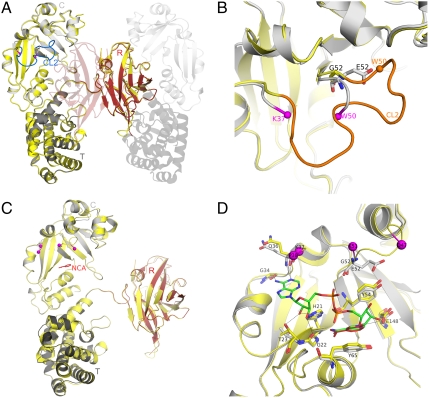
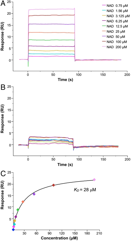
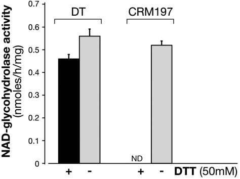
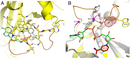
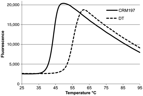
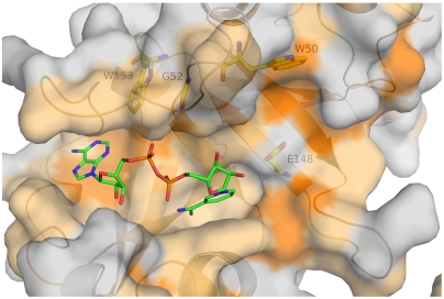
CRM197 is an enzymatically inactive and nontoxic form of diphtheria toxin that contains a single amino acid substitution (G52E). Being naturally nontoxic, CRM197 is an ideal carrier protein for conjugate vaccines against encapsulated bacteria and is currently used to vaccinate children globally against Haemophilus influenzae, pneumococcus, and meningococcus. To understand the molecular basis for lack of toxicity in CRM197, we determined the crystal structures of the full-length nucleotide-free CRM197 and of CRM197 in complex with the NAD hydrolysis product nicotinamide (NCA), both at 2.0-Å resolution. The structures show for the first time that the overall fold of CRM197 and DT are nearly identical and that the striking functional difference between the two proteins can be explained by a flexible active-site loop that covers the NAD binding pocket. We present the molecular basis for the increased flexibility of the active-site loop in CRM197 as unveiled by molecular dynamics simulations. These structural insights, combined with surface plasmon resonance, NAD hydrolysis, and differential scanning fluorimetry data, contribute to a comprehensive characterization of the vaccine carrier protein, CRM197.


Conflict of interest statement

Conflict of interest statement: The sponsor is a full-time employee of Novartis Vaccines

Figures

Fig. 1.
Fig. 1.
NF- and NCA-CRM197. (A) CRM197 catalytic (C) (residues 1–187), transmembrane (T) (residues 201–384), and receptor (R) (residues 387–535) domains are colored as light gray, dark gray, and red, respectively, and labeled. Nucleotide-free DT (PDB ID code 1SGK) is shown as yellow cartoon after superposition onto nucleotide-free CRM197. The active-site loop (CL2) of DT is shown in blue. The symmetry-related, domain-swapped molecule that forms the dimer of CRM197 is depicted using transparent cartoons. (B) Zoom into the superimposed catalytic domain only, showing the active-site loop (CL2) from DT in orange, the position of E52 of CRM197 and G52 of DT as sticks with carbon atoms colored in gray and yellow, respectively. The Cα atoms of the last visible residues of the disordered active-site loop of CRM197 (K37 and W50) are shown as spheres and colored in magenta. Nitrogen and oxygen atoms are colored in blue and red, respectively. (C) NAD-bound DT (PDB ID code 1TOX) is depicted as yellow cartoon after superposition onto NCA-CRM197 that is colored by domains as in Fig. 1A. NCA bound to CRM197 is shown as red sticks and labeled, and the last visible residues of the active-site loop of CRM197 and DT complexed with NAD are shown as spheres and colored in magenta. (D) The NAD-binding site in CRM197 (gray) compared to DT (yellow). Residues known to be involved both in NAD-binding (G22, T23, G34, Q36, Y54, Y65) or in catalysis (H21, E148) are shown as sticks for DT (carbon atoms in yellow) and CRM197 (carbon atoms in gray). E52 and G52 in CRM197 and DT are shown as sticks, while last visible residues of the active-site loop in both NCA-CRM197 (K37, K51) and NAD-DT (P38, D47) are shown as spheres and colored magenta. The NAD bound to DT is shown as sticks with carbon atoms colored in green, and nitrogen, oxygen, and phosphorous atoms, colored in blue, red, and orange, respectively. The NCA bound to CRM197 is shown as red sticks and labeled.
Fig. 2.
Fig. 2.
CRM197 has residual affinity for NAD. Increase of resonance units upon binding of NAD to immobilized DT and CRM197 is shown in A and B, respectively. The plateau line represents the steady-state equilibrium phase of interactions between the proteins and NAD, whereas the decrease in resonance from the plateau represents the dissociation phase. (C) Steady-state analysis of NAD binding to DT.
Fig. 3.
Fig. 3.
CRM197 in nonreducing conditions has NAD-glycohydrolase activity. The NAD-glycohydrolase activity of DT (20 μg) and CRM197 (20 μg) was carried out in 50 mM potassium phosphate, pH 7.5, 0.1 mM [carbonyl-14C]NAD (0.05 μCi) in a total volume of 0.15 ml. The activity was measured in the presence (black bars) or in the absence (gray bars) of 50 mM DTT. Samples (50 μL) after incubation at 37 C for 18 h were applied to 1-mL column of Dowex AG 1-X2 and 14C-nicotinamide eluted with 5 mL of H2O for liquid scintillation counting. NAD-glycohydrolase is expressed as nmol/h/mg and the incorporated radioactivity was measured in a Packard Top counter. ND, not detectable. Data are the means SD of values from two independent experiments.
Fig. 4.
Fig. 4.
Active-site loop conformation of CRM197 sampled by MD. (A) The active-site loop of NF-DT (PDB ID code 1SGK) is shown in orange. Residues of the loop involved in hydrogen bonds (P38, T42, K51, W153) are shown in sticks with carbon atoms colored in yellow, and the H-bond shown with dashes. Residues of the active site (I35, K37, F53) contributing to a hydrophobic environment around W153 are also shown as sticks. As reference, the NAD from the complex NAD-DT (PDB ID code 1TOX) is superimposed on NF-DT and shown as sticks in the background with carbon atoms colored in green. (B) Reoriented view of the active site. NF-CRM197 and NCA-CRM197 are shown in gray, after superposition on NF-DT (yellow). W50, G52, E52, and W153 from NF-DT, and from NCA-CRM197 and NF-CRM197 are shown in sticks with carbon atoms colored in yellow and gray for DT and CRM197, respectively. OE1 and OE2 atoms of E52 of CRM197 are shown as dots.
Fig. 5.
Fig. 5.
CRM197 is less thermostable than DT. Thermal denaturation was induced heating CRM197 and DT from 25 °C to 95 °C. CRM197 and DT displayed Tm values of 44.3 °C and 55.2 °C respectively and 10.9 °C ΔTm , indicating a greater stability of DT in comparison to CRM197.
Fig. 6.
Fig. 6.
Hydrophobicity of the NAD-binding pocket of DT. NF-DT (1SGK) is shown as gray surface and hydrophobic residues are colored using a scale from light to bright orange. The superimposed NAD molecule from the structure NAD-DT (PDB ID code 1TOX) is shown as sticks with carbon, oxygen, nitrogen, and phosphorous atoms colored in green, red, blue, and orange, respectively. Residues W153, G52, W50, and E148 are shown as sticks under the transparent surface, with carbon, oxygen, and nitrogen atoms colored in yellow, red, and blue, respectively.

References

    1. Hadfield TL, McEvoy P, Polotsky Y, Tzinserling VA, Yakovlev AA. The pathology of diphtheria. J Infect Dis. 2000;181:S116–S120. - PubMed
    1. Centers for Disease Control and Prevention. Diphtheria epidemic—new independent states of the former Soviet Union, 1990–1994. MMWR Morb Mortal Wkly Rep. 1995;44:177–181. - PubMed
    1. Pappenheimer AM, Gill DM. Diphtheria. Science. 1973;182:353–358. - PubMed
    1. Roux E, Yersin A. Contribution a l’etude de la diphthérie [Contribution to the study of diphtheria] Ann Inst Pasteur (Paris) 1888;2:629–661. in French.
    1. FitzGerald JG. Diphtheria toxoid as an immunizing agent. Can Med Assoc J. 1927;17:524–529. - PMC - PubMed

Publication types

LinkOut - more resources