. 2012 Apr 3;109(14):5499-504.
doi: 10.1073/pnas.1201132109. Epub 2012 Mar 19.

TWIK1, a unique background channel with variable ion selectivity

Affiliations

TWIK1, a unique background channel with variable ion selectivity

Franck C Chatelain et al. Proc Natl Acad Sci U S A. .

Abstract

TWIK1 belongs to the family of background K(+) channels with two pore domains. In native and transfected cells, TWIK1 is detected mainly in recycling endosomes. In principal cells in the kidney, TWIK1 gene inactivation leads to the loss of a nonselective cationic conductance, an unexpected effect that was attributed to adaptive regulation of other channels. Here, we show that TWIK1 ion selectivity is modulated by extracellular pH. Although TWIK1 is K(+) selective at neutral pH, it becomes permeable to Na(+) at the acidic pH found in endosomes. Selectivity recovery is slow after restoration of a neutral pH. Such hysteresis makes plausible a role of TWIK1 as a background channel in which selectivity and resulting inhibitory or excitatory influences on cell excitability rely on its recycling rate between internal acidic stores and the plasma membrane. TWIK1(-/-) pancreatic β cells are more polarized than control cells, confirming a depolarizing role of TWIK1 in kidney and pancreatic cells.


Conflict of interest statement

The authors declare no conflict of interest.

Figures

Fig. 1.
Fig. 1.
TWIK1 sequence and topological organization. (A) Functional domains in a TWIK1 subunit: transmembrane segments (M1–M4), coiled-coiled self-interacting domain (SID), pore domains (P1 and P2), pore helices (ph), and selectivity filter sequences (purple bold line) are indicated. (B) Sequence alignment in which TWIK1 residues mutated in this article. Residues affecting ion selectivity are in red; residues affecting channel activity are in blue. (C and D) Structural modeling of a TWIK1 dimer. Cytoplasmic and extracellular domains are not depicted. (C) Top view of the TWIK1 extracellular side. (D) Side views of the TWIK1 membrane domain; out-of-plane loops and helices are not depicted. Mutated residues shown in B are depicted.
Fig. 2.
Fig. 2.
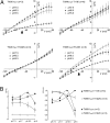
Pore mutations stimulate TWIK1-channel activity. (A) Currents evoked during voltage pulses ranging from −120 mV to +60 mV in 10-mV steps from a holding potential of −80 mV. Recordings were obtained from Xenopus oocytes 3 d after cRNA injection for TWIK1 and TWIK1I293A,I294A and 1 d after cRNA injection for TWIK1I293A,I294A,L146D. (B) I–V relationships deduced from currents after 1 d of expression. Each value is the mean ± SEM; n = 8 cells for TWIK1; n = 7 cells forTWIK1I293A,I294A; n = 7cells for TWIK1I293A,I294A,L146D. (C) After recording, oocytes in B were solubilized, and lysates were spotted in triplicate on membrane before detection by anti-TWIK1 antibodies. (D) I–V relationships deduced from currents after 1 d of expression. Each value is the mean ± SEM; n = 7 cells for TWIK1L146D; n = 8 cells for TWIK1L146D,K274E; n = 10 cells for TWIK1I293A,I294AL146D; n = 13 cells for TWIK1I293A,I294A,L146D,K274E. (E) The same experiment as in C but using oocytes recorded in D.
Fig. 3.
Fig. 3.
TWIK1 sensitivity to pH variations. (A) Effect of progressive extracellular acidification. For I–V relationships, each value is the mean ± SEM (number of cells is indicated for each condition). The shift of the Erev at pH 5.5 is indicated by an arrow. (B) Current variations at 0 mV. Each value is the mean ± SEM (number of cells is indicated for each condition).
Fig. 4.
Fig. 4.
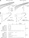
Ion selectivity of TWIK1 is altered upon acidification. (A) I–V relationships obtained in different external K+ conditions at pH 7.4 and 6. Na+ is replaced by K+. Each value is the mean ± SEM (n = 6 cells). (B) Erev as a function of the extracellular K+ concentration at pH 7.4 and 6. Each value is the mean ± SEM (n = 4 cells). (C) Relative PNa/PK of different TWIK1 mutants at pH 7.4 and 6. Each value is the mean ± SEM (n is indicated for each condition).
Fig. 5.
Fig. 5.
Kinetics of the ion selectivity change. (A) Effect of changing extracellular pH on TWIK1 currents recorded at −80 mV at 10-s intervals. (B) Effect of changing extracellular pH on TWIK1 current Erev recorded at 10-s intervals.
Fig. 6.
Fig. 6.
Depolarizing influence of TWIK1 in pancreatic β cells. (A) In pancreatic islets, TWIK1 is immunodetected in insulin-expressing B cells. Cells that do not express TWIK1 are glucagon- or somatostatin-positive cells. (Scale bar, 5 μM.) (B) Membrane potential (Em) of β cells primary-cultured from TWIK1−/− or control (WT) mice in various external glucose concentrations.

References

    1. Bayliss DA, Barrett PQ. Emerging roles for two-pore-domain potassium channels and their potential therapeutic impact. Trends Pharmacol Sci. 2008;29:566–575. - PMC - PubMed
    1. Goldstein SA, et al. International Union of Pharmacology. LV. Nomenclature and molecular relationships of two-P potassium channels. Pharmacol Rev. 2005;57:527–540. - PubMed
    1. Lesage F, Barhanin J. Molecular physiology of pH-sensitive background K(2P) channels. Physiology (Bethesda) 2011;26:424–437. - PubMed
    1. Noël J, Sandoz G, Lesage F. Molecular regulations governing TREK and TRAAK channel functions. Channels (Austin) 2011;5:402–409. - PMC - PubMed
    1. Arrighi I, Lesage F, Scimeca JC, Carle GF, Barhanin J. Structure, chromosome localization, and tissue distribution of the mouse twik K+ channel gene. FEBS Lett. 1998;425:310–316. - PubMed

Publication types

Substances