. 2012 May 11;287(20):16324-34.
doi: 10.1074/jbc.M112.342493. Epub 2012 Mar 19.

Structural rearrangements of the central region of the morbillivirus attachment protein stalk domain trigger F protein refolding for membrane fusion

Affiliations

Structural rearrangements of the central region of the morbillivirus attachment protein stalk domain trigger F protein refolding for membrane fusion

Nadine Ader et al. J Biol Chem. .

Abstract

It is unknown how receptor binding by the paramyxovirus attachment proteins (HN, H, or G) triggers the fusion (F) protein to fuse with the plasma membrane for cell entry. H-proteins of the morbillivirus genus consist of a stalk ectodomain supporting a cuboidal head; physiological oligomers consist of non-covalent dimer-of-dimers. We report here the successful engineering of intermolecular disulfide bonds within the central region (residues 91-115) of the morbillivirus H-stalk; a sub-domain that also encompasses the putative F-contacting section (residues 111-118). Remarkably, several intersubunit crosslinks abrogated membrane fusion, but bioactivity was restored under reducing conditions. This phenotype extended equally to H proteins derived from virulent and attenuated morbillivirus strains and was independent of the nature of the contacted receptor. Our data reveal that the morbillivirus H-stalk domain is composed of four tightly-packed subunits. Upon receptor binding, these subunits structurally rearrange, possibly inducing conformational changes within the central region of the stalk, which, in turn, promote fusion. Given that the fundamental architecture appears conserved among paramyxovirus attachment protein stalk domains, we predict that these motions may act as a universal paramyxovirus F-triggering mechanism.


Figures

FIGURE 1.
FIGURE 1.
The central section of the morbillivirus H-stalk is highly conserved. A, model of H-stalk motion upon receptor binding: a pre-formed tetrameric H-stalk (close) dissociates after receptor interaction (open). Each monomer of the H-tetramer is highlighted with different colors. B, upper panel: primary structure of the H-protein; CT, cytoplasmic tail; TM, transmembrane region; β1-β6, color-coded β-propeller blades 1–6. The four β-strands of blade 6 are indicated. Lower panel: sequence alignment of the amino acids of the H-proteins' stalk domain of different morbilliviruses. GenBankTM numbers for each virus sequence are: AB016162.1 (measles, ICB strain), AF266288.1 (measles, Edmonston strain), AF305419.1 (canine distemper, Onderstepoort strain), AY386316.1 (canine distemper, A75/17 strain), X98291.3 (rinderpest virus, Kabete O strain), Y18816.1 (rinderpest virus, K strain), FJ648457.1 (porpoise morbillvirus, IRL88 strain), FJ648456.1 (phocine distemper virus, DK02 strain), NC_005283.1 (dolphin morbillivirus). Red, blue, and black colors represent highly conserved, semi-conserved, and less conserved residues, respectively (15). Cysteines 139 and 154 at the top of the Morbillivirus H-stalk domain involved in natural disulfide bonds are shown (boxes). C, side view of the PIV5 HN-stalk structure. The upper 4HB straight structure is shown in green, the lower superhelical region in purple. D, structural model of the CDV H-stalk (side view). The region subjected to cysteine-scanning mutagenesis is highlighted in red. Putative F-contacting region (111–118) is shown in blue (23).
FIGURE 2.
FIGURE 2.
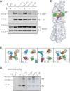
Successful engineering of intermonomers disulfide bonds. A, assessment of covalent H-tetramers. Surface-exposed material (surface immunoprecipitation; anti-FLAG mAb) of Vero cells transfected with plasmids encoding the various FLAG-tagged H-constructs, or empty plasmids (pCI) were analyzed. Precipitates were boiled and fractionated in Tris/acetate gels under nonreducing conditions, H-antigenic material was detected with a polyclonal anti-H antibody (41). H4: H-tetramers; H2: H-dimers, H1: H-monomers. B, assessment of disulfide bond formation in an H-C139A/C154A cysteine-free background. Vero cells transfected with plasmids encoding selected H mutants were lysed with stringent RIPA buffer. Lysates were boiled and fractionated in Tris/glycine gels under reducing and nonreducing conditions. H4: H-tetramers; H2: H-dimers, H1: H-monomers. C, assessment of disulfide bond generation in soluble H. Supernatants of transfected 293T cells were harvested 3 days post-transfection. Soluble H-proteins were then purified using nickel-based magnetic beads and 1 μg of protein was then fractionated either in Tris/acetate gels (left panel) or in native gels (right panel). H-antigenic material was detected with the polyclonal anti-H antibody. H4: H-tetramers; H2: H-dimers, H1: H-monomers.
FIGURE 3.
FIGURE 3.
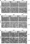
Reversible inhibition of F/H-induced cell-to-cell fusion by mild DTT-treatment. A, upper panel: graphic representation of fusion assay results, obtained in receptor-positive cells (Vero-cSLAM). Each bar represents a single cysteine substitution in the H-stalk sequence (lower panel; representation of H as in Fig. 1B). For fusion assays, Vero-cSLAM cells were transfected with plasmids encoding the F and H genes, followed by treatment overnight with a fusion-inhibiting molecule (36, 37). Fusion activity was analyzed 24 h post-transfection, after DTT treatment (30 min, 15 mm: +DTT), or without further treatment (−DTT). Each bar is partitioned in 3 parts, corresponding to three different levels of fusion-promotion activity of the different H mutants (0: no syncytium formation in 5 representative fields of views; 1: limited number of syncytia monitored in 5 representative fields of views; 2: intermediate numbers of syncytia monitored in 5 representative fields of views; 3: massive numbers of syncytia monitored in 5 representative fields of views). An average fusion score was assigned to each mutant based on three independent experiments. Bars filled with dark green: fusion activity promoted by the different H mutants; bars filled with light green, restored (or boosted) fusion activity after DTT-treatment. CS, crystal structure. B, upper panels: CDV H 4HB-stalk model; predicted positions of H-residue side chains of constructs that were selected for the quantitative fusion assays. Each monomer is color-coded. Lower panels: syncytium formation after co-transfection of Vero-cSLAM cells with plasmid DNA encoding various CDV-H proteins and F. Twenty-four hours post-transfection, cells were treated with DTT (15 mm) or left untreated; representative fields of view are shown. C, quantitation of F-triggering activity of selected H mutants using a luciferase reporter gene-based content mix assay. Vero-SLAM cells were infected with MVA-T7 (MOI of 1). In parallel, a second population (Vero cells) was transfected with plasmids encoding the different H proteins, a plasmid encoding F and a plasmid containing the luciferase reporter gene under the control of the T7 promoter. Twenty-four hours post-transfection, Vero cells were mixed with Vero-cSLAM cells and seeded into fresh plates. Cells were then incubated for 2 h in the presence of the fusion-inhibition molecule (3g), followed by extensive washing and incubation with or without DTT (15 mm) for 30 min. After 3 h at 37 °C, fusion was quantified using a commercial luciferase-measuring kit. For each experiment, luciferase activity of the Fwt/Hwt combination was set to 100%. Means and standard deviations of three independent experiments, performed in duplicate, are shown. Dark gray histograms: F/H-mediated fusion in the absence of DTT treatment; Light gray histograms: F/H-mediated fusion in the presence of DTT treatment.
FIGURE 4.
FIGURE 4.
Structural flexibility of the central morbillivirus H-stalk region is universally required to trigger fusion. Syncytium formation after co-transfection of cells with plasmid DNA encoding various morbillivirus H and F proteins. After transfection, the cells were treated overnight with a fusion-inhibition molecule (3g for CDV and FIP for MeV). Twenty-four hours post-transfection, cells were washed and subsequently treated with DTT (15 mm) or left untreated; representative fields of view are shown. A, transfections were performed with CDV F and H-expression plasmids in Vero-cSLAM cells. B, transfections were performed with MeV F and H-expression plasmids in Vero-hSLAM cells. C, transfections were performed with MeV F and H-expression plasmids in Vero cells.
FIGURE 5.
FIGURE 5.
The 111–114 section of the CDV H-stalk domain regulates F binding activity. A, assessment of H interaction with functional F proteins. To stabilize the F/H interactions, Vero cells co-transfected with F- and H-expression plasmids (or empty plasmid: pCI) were treated with the membrane-impermeable crosslinker DTSSP. Subsequently, Vero cells were lysed with stringent RIPA buffer, followed by H immunoprecipitation (IP) with an anti-H mAb (1347) recognizing a linear epitope and protein G-Sepharose beads. Precipitates were boiled and subjected to immunoblotting using a polyclonal anti-F antiserum for F detection (41). In addition, F-proteins present in the lysates prior to IP were detected by immunoblot using a polyclonal anti-F antibody (TL). As a control, total H immunoprecipitates, were analyzed by immunoblotting using a polyclonal anti-H antibody (IP). H1: H-monomers; F0: uncleaved F precursors; F1: cleaved F precursors. B, CDV H 4HB-stalk model; the side chains of residues H-L111, H-N112, H-E113, and H-I114 are highlighted, and each residue is color-coded. Inset: fusion-triggering capacity of each H mutant (supplemental Fig. S3). C, surface representation of the structural model of the CDV H-stalk (bottom is membrane-proximal). Positions of residues H-L111 (in red), H-E113 (in green), and H-I114 (in yellow) are shown. D, assessment of covalent tetramerization of H variants harboring cysteine residues at consecutive positions from residue 111 to 114. Cell surface-exposed material was analyzed as detailed in Fig. 2A.
FIGURE 6.
FIGURE 6.
Model of paramyxovirus attachment protein-mediated F-triggering activity. A, prior to receptor binding, the tightly packed tetrameric attachment protein stalk domain, which supports the four receptor-binding heads, sustains physical interaction with F. B, upon receptor binding, the heads rearrange, in turn applying forces to the stalk and the transmembrane domains. Because SLAM is thought to bind to the H-head laterally (9), this may result in reducing the distance between target and effector membranes (initial position of the target membrane is represented by a dashed white line). C, putative metastable supercoiled lower section of the stalk then unwinds, thereby inducing intersubunits rearrangements of the central stalk region, which is predicted to contact F. This central section, and possibly the conserved proline residue at position 108, is then pivotal in F-triggering. The red star indicates active H-mediated F-triggering. D, activated F undergoes structural rearrangements, reaching the pre-hairpin state. E, upper section: sequence alignment of a partial section of the attachment protein stalk domains of several paramyxoviruses (NDV, PIV3 and 5, MeV and CDV). Yellow: conserved residues; light blue: residues identified in CDV H that are DTT-sensitive when mutated to cysteine (fusion activity restored or boosted). Light red: residues in CDV and MeV H (23) identified as contributors to F interaction. Lower section: representation of the segment targeted by cysteine-scanning mutagenesis. Individual mutants and the corresponding F-triggering capacities are shown above: light green color indicates DTT-dependent and/or independent fusion promotion; dark red color indicates strong deficiency in fusion promotion (both in the absence and presence of DTT treatment).

References

    1. Chen S. Y., Anderson S., Kutty P. K., Lugo F., McDonald M., Rota P. A., Ortega-Sanchez I. R., Komatsu K., Armstrong G. L., Sunenshine R., Seward J. F. (2011) Health care-associated measles outbreak in the United States after an importation: challenges and economic impact. J. Infect. Dis. 203, 1517–1525 - PubMed
    1. Tatsuo H., Ono N., Tanaka K., Yanagi Y. (2000) SLAM (CDw150) is a cellular receptor for measles virus. Nature 406, 893–897 - PubMed
    1. Tatsuo H., Ono N., Yanagi Y. (2001) Morbilliviruses use signaling lymphocyte activation molecules (CD150) as cellular receptors. J. Virol. 75, 5842–5850 - PMC - PubMed
    1. Dörig R. E., Marcil A., Chopra A., Richardson C. D. (1993) The human CD46 molecule is a receptor for measles virus (Edmonston strain). Cell 75, 295–305 - PubMed
    1. Negrete O. A., Levroney E. L., Aguilar H. C., Bertolotti-Ciarlet A., Nazarian R., Tajyar S., Lee B. (2005) EphrinB2 is the entry receptor for Nipah virus, an emergent deadly paramyxovirus. Nature 436, 401–405 - PubMed

Publication types

Substances

LinkOut - more resources