Outbreaks of Pneumocystis pneumonia in 2 renal transplant centers linked to a single strain of Pneumocystis: implications for transmission and virulence
- PMID: 22431811
- PMCID: PMC3404729
- DOI: 10.1093/cid/cis217
Outbreaks of Pneumocystis pneumonia in 2 renal transplant centers linked to a single strain of Pneumocystis: implications for transmission and virulence
Abstract
Background: There have been numerous reports of clustered outbreaks of Pneumocystis pneumonia (PCP) at renal transplant centers over the past 2 decades. It has been unclear whether these outbreaks were linked epidemiologically to 1 or several unique strains, which could have implications for transmission patterns or strain virulence.
Methods: Restriction fragment length polymorphism (RFLP) analysis was used to compare Pneumocystis isolates from 3 outbreaks of PCP in renal transplant patients in Germany, Switzerland, and Japan, as well as nontransplant isolates from both human immunodeficiency virus (HIV)-infected and uninfected patients.
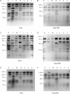
Results: Based on RFLP analysis, a single Pneumocystis strain caused pneumonia in transplant patients in Switzerland (7 patients) and Germany (14 patients). This strain was different from the strain that caused an outbreak in transplant patients in Japan, as well as strains causing sporadic cases of PCP in nontransplant patients with or without HIV infection.
Conclusions: Two geographically distinct clusters of PCP in Europe were due to a single strain of Pneumocystis. This suggests either enhanced virulence of this strain in transplant patients or a common, but unidentified, source of transmission. Outbreaks of PCP can be better understood by enhanced knowledge of transmission patterns and strain variation.
Figures


Comment in
-
Linking Pneumocystis epidemiology, transmission, and virulence.Clin Infect Dis. 2012 May;54(10):1445-7. doi: 10.1093/cid/cis216. Epub 2012 Mar 19. Clin Infect Dis. 2012. PMID: 22431810 No abstract available.
-
Pneumocystis jirovecii genotypes involved in pneumocystis pneumonia outbreaks among renal transplant recipients.Clin Infect Dis. 2013 Jan;56(1):165-6. doi: 10.1093/cid/cis810. Epub 2012 Sep 18. Clin Infect Dis. 2013. PMID: 22990847 No abstract available.
-
Reply to Hauser et Al.Clin Infect Dis. 2013 Jan;56(1):166-7. doi: 10.1093/cid/cis814. Epub 2012 Sep 18. Clin Infect Dis. 2013. PMID: 22990850 Free PMC article. No abstract available.
References
-
- Kovacs JA, Masur H. Evolving health effects of Pneumocystis: one hundred years of progress in diagnosis and treatment. JAMA. 2009;301:2578–85. - PubMed
-
- Krajicek BJ, Limper AH, Thomas CF., Jr Advances in the biology, pathogenesis and identification of Pneumocystis pneumonia. Curr Opin Pulm Med. 2008;14:228–34. - PubMed
-
- Arichi N, Kishikawa H, Mitsui Y, et al. Cluster outbreak of Pneumocystis pneumonia among kidney transplant patients within a single center. Transplant Proc. 2009;41:170–2. - PubMed
-
- de Boer MG, Bruijnesteijn van Coppenraet LE, Gaasbeek A, et al. An outbreak of Pneumocystis jiroveci pneumonia with 1 predominant genotype among renal transplant recipients: interhuman transmission or a common environmental source? Clin Infect Dis. 2007;44:1143–9. - PubMed
-
- Gianella S, Haeberli L, Joos B, et al. Molecular evidence of interhuman transmission in an outbreak of Pneumocystis jirovecii pneumonia among renal transplant recipients. Transpl Infect Dis. 2010;12:1–10. - PubMed
Publication types
MeSH terms
Grants and funding
LinkOut - more resources
Full Text Sources
Medical
Miscellaneous

