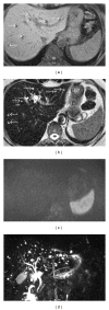
Computed tomography and magnetic resonance imaging findings in a case with biliary microhamartomas
- PMID: 22431945
- PMCID: PMC3295846
- DOI: 10.1155/2012/976078
Computed tomography and magnetic resonance imaging findings in a case with biliary microhamartomas
Abstract
Biliary microhamartomas, also known as bile duct hamartomas and von Meyenburg complexes, are benign neoplasms containing cystic dilated bile ducts embedded in fibrous stroma. They develop in hepatobiliary system, do not generally give clinical outcomes, and are detected incidentally. However, they can rarely show malignant transformation. Our aim was to report the contribution of computed tomography, routine magnetic resonance imaging, and magnetic resonance cholangiopancreatography in the diagnosis of biliary microhamartomas in a 61-year-old woman.
Figures


References
-
- Dahnert W. Liver, bile ducts, pancreas and spleen. In: Dahnert W, editor. Radiology Review Manual. 6th edition. Philadelphia, Pa, USA: Lippincott Williams and Wilkins; 2007. pp. 732–733.
-
- Xu AM, Xian ZH, Zhang SH, Chen XF. Intrahepatic cholangiocarcinoma arising in multiple bile duct hamartomas: report of two cases and review of the literature. European Journal of Gastroenterology and Hepatology. 2009;21(5):580–584. - PubMed
-
- Redston MS, Wanless IR. The hepatic von Meyenburg complex: prevalence and association with hepatic and renal cysts among 2843 autopsies [corrected] Modern Pathology. 1996;9(3):233–237. - PubMed
-
- Thommesen N. Biliary hamartomas (von Meyenburg complexes) in liver needle biopsies. Acta Pathologica et Microbiologica Scandinavica. 1978;86(2):93–99. - PubMed
-
- Schneider G, Grazioli L, Saini S. Imaging of the biliary tree and gallbladder diseaese. In: Schneider G, Grazioli L, Saini S, editors. MRI of the Liver. 2nd edition. Milan, Italy: Springer; 2006. p. 252.
Publication types
LinkOut - more resources
Full Text Sources

