. 2012;7(3):e33266.
doi: 10.1371/journal.pone.0033266. Epub 2012 Mar 14.

ER stress in retinal degeneration in S334ter Rho rats

Affiliations

ER stress in retinal degeneration in S334ter Rho rats

Vishal M Shinde et al. PLoS One. 2012.

Abstract

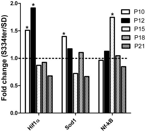
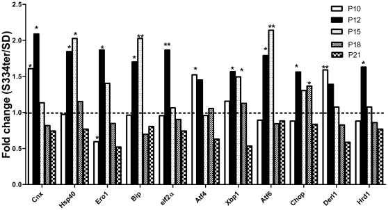
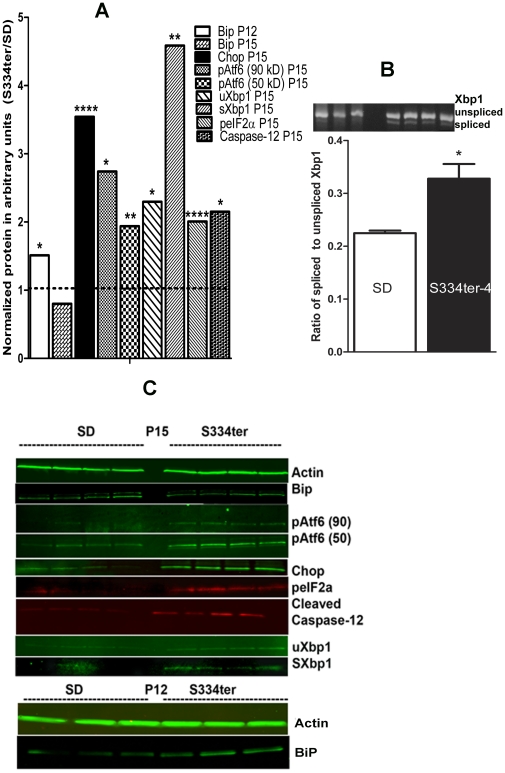
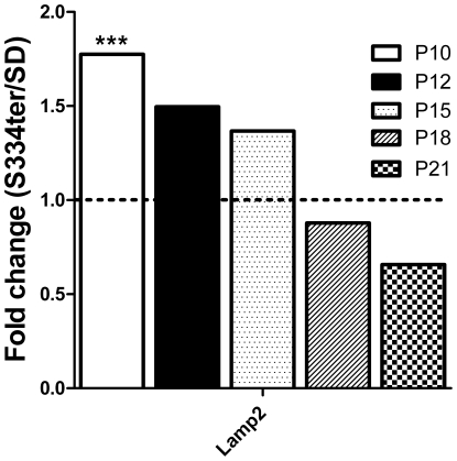
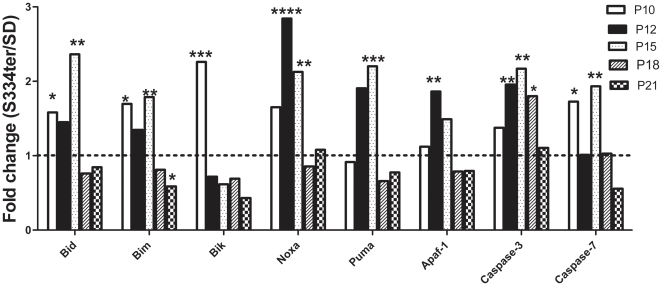
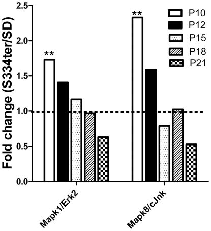
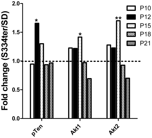
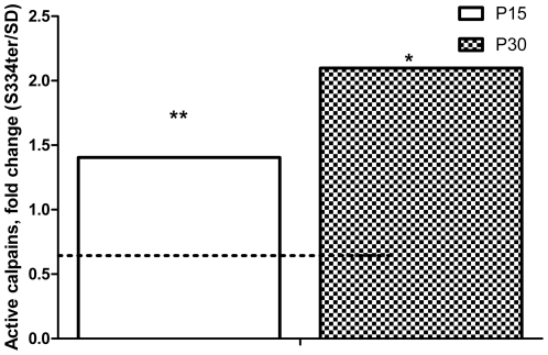
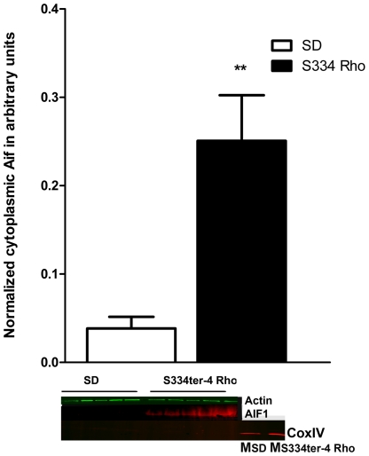
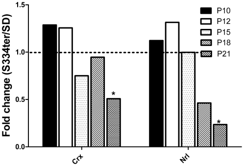
The S334ter rhodopsin (Rho) rat (line 4) bears the rhodopsin gene with an early termination codon at residue 334 that is a model for several such mutations found in human patients with autosomal dominant retinitis pigmentosa (ADRP). The Unfolded Protein Response (UPR) is implicated in the pathophysiology of several retinal disorders including ADRP in P23H Rho rats. The aim of this study was to examine the onset of UPR gene expression in S334ter Rho retinas to determine if UPR is activated in ADRP animal models and to investigate how the activation of UPR molecules leads to the final demise of S334ter Rho photoreceptors. RT-PCR was performed to evaluate the gene expression profiles for the P10, P12, P15, and P21 stages of the development and progression of ADRP in S334ter Rho photoreceptors. We determined that during the P12-P15 period, ER stress-related genes are strongly upregulated in transgenic retinas, resulting in the activation of the UPR that was confirmed using western blot analysis and RT-PCR. The activation of UPR was associated with the increased expression of JNK, Bik, Bim, Bid, Noxa, and Puma genes and cleavage of caspase-12 that together with activated calpains presumably compromise the integrity of the mitochondrial MPTP, leading to the release of pro-apoptotic AIF1 into the cytosol of S334ter Rho photoreceptor cells. Therefore, two major cross-talking pathways, the UPR and mitochondrial MPTP occur in S334ter-4 Rho retina concomitantly and eventually promote the death of the photoreceptor cells.


Conflict of interest statement

Competing Interests: The authors have declared that no competing interests exist.

Figures

Figure 1
Figure 1. Relative expression of the oxygen stress-induced Hif1a, Sod1 and Nf-kB genes in S334ter-4 Rho retinas at different ages.
Relative gene expression in S334ter-4 Rho retina was measured on P10, P12, P15, P18 and P21 and a fold change was expressed as a ratio of S334ter-4Rho relative expression to SD relative expression. Expression of the Hif1α and Sod1 genes was significantly induced in P10 S334ter-4 Rho compared to SD retinas (1.5- and 1.4-fold, respectively, P<0.05, *). On P12, increased Hif1a expression was still evident (1.9-fold, P<0.05,*); however, the Sod1 expression was decreased compared to P10. When compared with Hif1a and Sod1 mRNA, the relative accumulation of Nf-kB mRNA in S334ter-4 Rho retinas exhibited alternate dynamics: a 1.7-fold increase in Nf-kB expression was observed on P15 only (P<0.05,*), whereas the Hif1α and Sod1 expression was diminished on P15.
Figure 2
Figure 2. Relative expression of ER stress- and ERAD-related genes in S334ter-4 Rho retinas.
The UPR gene expression was altered in S334ter-4 RHO retinas. Relative gene expression in S334tr Rho retina was measured on P10, P12, P15, P18 and P21 and a fold change was expressed as a ratio of S334ter-4Rho relative expression to SD relative expression. On P10, there was a significant reduction in the Ero1 gene expression suggesting that the ER homeostasis in S334ter-4 Rho retinas is imbalanced. The expression of this gene is decreased 2-fold (P<0.05, *) in S334ter-4 Rho retinas compared with SD retinas on P10. The increased expression of Calnexin (Cnx) and Atf4 genes are the first ER stress markers that respond to ER disturbance. The expression of these genes was increased 1.6- and 1.5-fold, respectively, (P<0.05, * in each case) on P10. On P12, the expression of other UPR upstream and downstream markers, such as Hsp40/Dnajc10, Ero1, Bip, eIf2a, Xbp1, Atf6 and Chop, were detected, which was indicated by relative increases of 1.8-, 1.8-, 1.5-, 1.9-, 1.6-, 1.8- and 1.6-fold, respectively, (P<0.05, * in each case with the exception of eIF2a where P<0.01, **). On P15, the Cnx, Ero1, eIf2a, Atf4 and Chop genes were expressed to a lesser extent or there was no significant difference in their expression levels in S334ter-4 Rho retinas compared with SD retinas. However, the Hsp40/Dnajc10, Bip, Xbp1 and ATf6 mRNAs were significantly induced on P15. Their relative expressions were 2-, 2-, 1.5- and 2-fold higher in S334ter-4 Rho retinas compared with the WT group (P<0.01 for Bip and Atf6, and P<0.05 for Hsp40/Dnajc10 and Xbp1). On P21, the expression of all genes was insignificant in the S334ter-4 Rho retinas compared with the SD retinas. Derl1 and Hrd1 gene expression was upregulated 1.5- and 1.6-fold on p10 and p12, respectively (P<0.05 in each case).
Figure 3
Figure 3. The ER stress markers BIP and CHOP proteins in retinas from S334ter-4 Rho rats.
A: Ratios of normalized S334ter-4 Rho BiP, CHOP, pATF6 (90), pATF6 (50), peIF2α ,spliced Xbp1(sXbp1), unsliced Xbp1 (uXbp1), cleaved caspase-12 proteins to corresponding proteins in SD CHOP were used to register the alteration of protein expression. Normalization of all proteins was done by detecting β-Actin protein The ER stress markers BIP protein was a 1.5-fold upregulated in P12 S334ter-4 Rho retinas compared with SD retinas (0.047±0.008 vs. 0.071±0.005, respectively; P = 0.01). In P15, the level of BiP protein was significantly reduced and was not distinguishable from SD. The Chop protein was also dramatically over-expressed (3.5-fold) (0.016±0.005 in SD vs. 0.60±0.002 S334ter-4 Rho, P = 0.0003) on P15 in S334ter-4 Rho retinas. The full length of pAtf6 protein (90 kD) (the Atf6 pathway) was significantly elevated in S334ter-4 Rho retina by 2.7-fold (0.0017±0.0011 in SD vs. 0.0048±0.0007 in S334ter-4 Rho, P = 0.04). The cleaved pAtf6 (50) was significantly elevated by 1.93-fold inS334ter-4 Rho retina (0.18±0.004 in SD vs. 0.035±0.002, P = 0.004). The peIF2α protein was also significantly increased in S334ter-4 Rho retina and was 0.001±0.0005 in S334ter-4 Rho vs 0.002±0.0004, P = 0.0009. The full length of pAtf6 protein (90 kD) (the Atf6 pathway) was significantly elevated in S334ter-4 Rho retina by 2.7-fold (0.0017±0.0011 in SD vs. 0.0048±0.0007 in S334ter-4 Rho, P = 0.04). The N-terminal of the full-length of Atf6, cleaved pAtf6 was elevated by 1.93-fold and was 0.18±0.004 in SD vs. 0.035±0.002, P = 0.004. We also observed that the peIF2α protein was significantly increased in S334ter-4 Rho retina and was 0.001±0.0005 in S334ter-4 Rho vs 0.002±0.0004, P = 0.0009. The spliced Xbp1 protein was detected in S334ter-4 retina. Its level was a 4.5-fold higher in transgenic retina compared to SD and was 0.022±0.003 in SD and 0.1±0.01, P = 0.001 in S334ter-4 rats. Unspliced Xbp1 protein was increased to a lesser extent (2.3-fold) in S334ter-4 rats and was 0.049±0.008 in SD and 0.13±0.001, P = 0.034 in S334ter-4. Increase in active caspase-12 was observed in S334ter-4 Rho retina. The level of cleaved caspase-12 (20 kD) was elevated in S334ter-4 Rho rats on P15 compared to control over 2-fold and was 0.05±0.007 in SD vs. 0.12±0.02 in S334ter-4 Rho, P = 0.014 on P15. B: Upper panel: Quantification of spliced form of the Xbp1 mRNA (the IRE signaling) detected by RT-PCR reaction. We observed 1.45-fold increased in the spliced form of Xbp1 mRNA in S334ter-4 Rho retina. The ratio of spliced Xbp1 to normalized unspliced Xbp1 mRNA was 0.22±0.0006 in SD vs. 0.033±0.031 in S334ter-4 Rho retina, P = 0.025). Image of the agarose gel loaded with RT-PCR product obtained with Xbp1 specific primers is shown in a lower panel. C: Images of western blots treated with anti-Actin, Bip, CHOP, peIf2α, pATF6, Xbp1 antibodies and detected with secondary antibodies and infrared imaging scanner are presented.
Figure 4
Figure 4. Relative expression of Lamp2 in S334ter-4 Rho retinas.
Relative gene expression in S334tr Rho retina was measured on P10, P12, P15, P18 and P21 and a fold change was expressed as a ratio of S334ter-4Rho relative expression to SD relative expression. The Lamp2 gene expression was upregulated in p10-p15 S334ter-4 Rho retinas. In p10, the relative expression of this gene increased 1.7-fold in S334ter-4 Rho retinas compared to that in SD retinas (P<0.001), and it subsequently declined in a time-dependent manner.
Figure 5
Figure 5. Relative expression of pro-apoptotic genes involved in S334ter-4 Rho retinas.
Relative gene expression in S334ter-4 Rho retina was measured on P10, P12, P15, P18 and P21 and a fold change was expressed as a ratio of S334ter-4 Rho relative expression to SD relative expression. The expression of BH3-only proteins, BID, Bim, Bik, Noxa and Puma was upregulated with different patterns in S334ter-4 Rho retinas. On P10, the expression of Bik, Bim and Bid genes increased 2.3- (P<0.001) 1.7- (P<0.05) and 1.6-fold (P<0.05), respectively. The expression of Bim was elevated until p15 and dropped 0.6-fold to WT levels (P<0.05) on P21. On P12, the Noxa gene was elevated 2.8-fold (P<0.0001) and remained upregulated until p15 (2-fold increase, P<0.01) and its expression subsequently dropped. On P15, the Puma gene appears to be upregulated 2.2-fold in S334ter-4 Rho retinas when compared to SD retinas (P<0.001). The Apaf1 gene expression was increased in P12 and in P15 retinas. A significant 1.9-fold upregulation Apaf1 expression was detected on P12 (P<0.01). The relative expression of caspase-3 was significantly upregulated during the progression of ADRP. On P12, P15 and P18, caspase-3 expression in S334ter-4 Rho retinas was elevated 1.9-fold (P<0.01), 2.2-fold (P<0.01) and 1.8-fold (P<0.05), respectively. compared to SD retinas. The analysis of caspase-7 gene expression demonstrates a 1.7-fold (P<0.05) and 2-fold (P<0.01) increase in caspase-7 mRNA on P10 and P15, respectively.
Figure 6
Figure 6. Relative expression of the MAPK1 (Erk2) and MAPK8 (JNK) genes in S334ter-4 Rho retinas.
Relative gene expression in S334ter-4 Rho retina was measured on P10, P12, P15, P18 and P21 and a fold change was expressed as a ratio of S334ter-4Rho relative expression to SD relative expression. On P10, the Mapk3 (Erk1) and Mapk14 (p38) gene expression was upregulated 1.7- and 2.3-fold in S334ter-4 Rho retinas, respectively (P<0.01 for both). Starting on P12, their expression decreased with time.
Figure 7
Figure 7. Relative expression of pTten, Akt1 and Akt2 genes in S334ter-4 Rho retinas.
Relative gene expression in S334ter-4 Rho retina was measured on P10, P12, P15, P18 and P21 and a fold change was expressed as a ratio of S334ter-4Rho relative expression to SD relative expression. An approximate 1.65-fold increase in the relative expression of the pTen gene was observed (0.80±0.13 vs. 1.5±0.2, P<0.05) in P12 transgenic retinas. Subsequently, the expression of this gene decreased to WT levels. In contrast, the expression of the Akt1 and 2 genes increased after P12 and reached a peak on P15. The Akt1 gene expression was 0.84±0.15 in SD retinas vs. 1.40±0.13 in S334ter-4 Rho retinas, P<0.05. The Akt2 gene expression was 1.03±0.17 in SD vs. 1.70±0.15 in P15 S334ter-4 Rho, P<0.01.
Figure 8
Figure 8. Activation of calpains in S334ter-4 Rho retinas.
In P15 and P30 S334ter-4 Rho retinas, we observed an activation of calpains in the cytosol measured by the fluorescence intensity emitted from the calpain substrate Ac-LLY AFC. On P15 and P30, there was a 1.4- and a greater than 2-fold increase in activated calpains in S334ter-4 Rho retinas compared with SD retinas, respectively (2.24±0.09 in SD vs. 3.1±0.127 in S334ter-4 Rho, P<0.01 and 1.287±0.27 in SD vs. 2.7±0.04 in S334ter-4 Rho, P<0.05, respectively).
Figure 9
Figure 9. Release of AIF1 from S334ter-4 Rho mitochondria.
Analysis of isolated cytosolic fractions of P15 S334ter-4 Rho retinas determined that the concentration of the cleaved form of Aif1 (48 kD) was 6.5-fold higher compared with the WT cytosolic samples (0.038±0.01 in SD vs. 0.25±0.05 in S334ter-4 Rho, P = 0.004). Images of western blot detecting the AIF1 and COXIV proteins are shown. Detection of COXIV protein was observed only in SD (Msd) and S334ter-4 (MS s334ter-4) retinal mitochondrial fractions.
Figure 10
Figure 10. Relative expression of the Crx and Nrl transcription factors in S334ter-4 Rho retinas.
Relative gene expression in S334ter-4 Rho retina was measured on P10, P12, P15, P18 and P21 and a fold change was expressed as a ratio of S334ter-4Rho relative expression to SD relative expression. The qRT-PCR analysis of S334ter-4 Rho retinas revealed that the expression of the Nrl and Crx transcription factors decreased in a time-dependent manner in P10-P21 retinas. A significant reduction in the expression of both transcription factors was observed in P21 retinas, the Crx gene expression was reduced 55% (P<0.05), and the NRL gene expression was downregulated 23% (P<0.05).

Similar articles

Cited by

References

    1. Bowne SJ, Humphries MM, Sullivan LS, Kenna PF, Tam LC, et al. A dominant mutation in RPE65 identified by whole-exome sequencing causes retinitis pigmentosa with choroidal involvement. Eur J Hum Genet. 2011;19(10):1074–1081. - PMC - PubMed
    1. Hernan I, Gamundi MJ, Planas E, Borras E, Maseras M, et al. Cellular expression and siRNA-mediated interference of rhodopsin cis-acting splicing mutants associated with autosomal dominant retinitis pigmentosa. Invest Ophthalmol Vis Sci. 2011;52:3723–3729. - PubMed
    1. Lee EH, Wu C, Lee EU, Stoute A, Hanson H, et al. Fatalities associated with the 2009 H1N1 influenza A virus in New York city. Clin Infect Dis. 2010;50:1498–1504. - PubMed
    1. Green ES, Menz MD, LaVail MM, Flannery JG. Characterization of rhodopsin mis-sorting and constitutive activation in a transgenic rat model of retinitis pigmentosa. Invest Ophthalmol Vis Sci. 2000;41:1546–1553. - PubMed
    1. Travis GH. Mechanisms of cell death in the inherited retinal degenerations. Am J Hum Genet. 1998;62:503–508. - PMC - PubMed

Publication types

MeSH terms