

Object working memory performance depends on microstructure of the frontal-occipital fasciculus
- PMID: 22432421
- PMCID: PMC3319977
- DOI: 10.1089/brain.2011.0037
Object working memory performance depends on microstructure of the frontal-occipital fasciculus
Abstract
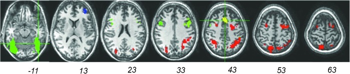
Re-entrant circuits involving communication between the frontal cortex and other brain areas have been hypothesized to be necessary for maintaining the sustained patterns of neural activity that represent information in working memory, but evidence has so far been indirect. If working memory maintenance indeed depends on such temporally precise and robust long-distance communication, then performance on a delayed recognition task should be highly dependent on the microstructural integrity of white-matter tracts connecting sensory areas with prefrontal cortex. This study explored the effect of variations in white-matter microstructure on working memory performance in two separate groups of participants: patients with multiple sclerosis and age- and sex-matched healthy adults. Functional magnetic resonance imaging was performed to reveal cortical regions involved in spatial and object working memory, which, in turn, were used to define specific frontal to extrastriate white-matter tracts of interest via diffusion tensor tractography. After factoring out variance due to age and the microstructure of a control tract (the corticospinal tract), the number of errors produced in the object working memory task was specifically related to the microstructure of the inferior frontal-occipital fasciculus. This result held for both groups, independently, providing a within-study replication with two different types of white-matter structural variability: multiple sclerosis-related damage and normal variation. The results demonstrate the importance of interactions between specific regions of the prefrontal cortex and sensory cortices for a nonspatial working memory task that preferentially activates those regions.
Figures


References
-
- Arnett PA. Rao SM. Bernardin L. Grafman J. Yetkin FZ. Lobeck L. Relationship between frontal lobe lesions and Wisconsin Card Sorting Test performance in patients with multiple sclerosis. Neurology. 1994;44:420–425. - PubMed
-
- Au Duong MV. Audoin B. Boulanouar K. Ibarrola D. Malikova I. Confort-Gouny S, et al. Altered functional connectivity related to white matter changes inside the working memory network at the very early stage of MS. J Cereb Blood Flow Metab. 2005a;25:1245–1253. - PubMed
-
- Au Duong MV. Boulanouar K. Audoin B. Terseras S. Ibarrola D. Malikova I, et al. Modulation of effective connectivity inside the working memory network in patients at the earliest stage of multiple sclerosis. Neuroimage. 2005b;24:533–538. - PubMed
-
- Bobholz JA. Rao SM. Cognitive dysfunction in multiple sclerosis: a review of recent developments. Curr Opin Neurol. 2003;16:283–238. - PubMed
-
- Bobholz JA. Rao SM. Lobeck L. Elsinger C. Gleason A. Kanz J, et al. fMRI study of episodic memory in relapsing-remitting MS: correlation with T2 lesion volume. Neurology. 2006;67:1640–1645. - PubMed
Publication types
MeSH terms
Grants and funding
LinkOut - more resources
Full Text Sources
