Review
. 2011;1(1):3-12.
doi: 10.1089/brain.2011.0019.

The restless brain

Affiliations
Review

The restless brain

Marcus E Raichle. Brain Connect. 2011.

Abstract

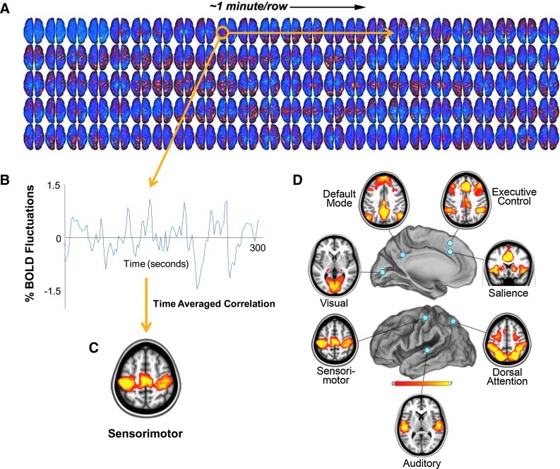
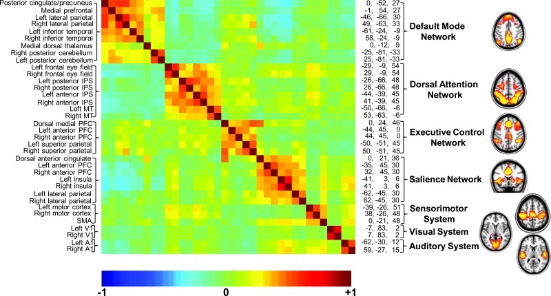
The pressing need to better understand human brain organization is appreciated by all who have labored to explain the uniqueness of human behavior in health and disease. Early work on the cytoarchitectonics of the human brain by Brodmann and others accompanied by several centuries of lesion behavior work, although valuable, has left us far short of what we need. Fortunately, modern brain imaging techniques have, over the past 40 years, substantially changed the situation by permitting the safe appraisal of both anatomical and functional relationships within the living human brain. An unexpected feature of this work is the critical importance of ongoing, intrinsic activity, which accounts for the majority of brain's energy consumption and exhibits a surprising level of organization that emerges with dimensions of both space and time. In this essay, some of the unique features of intrinsic activity are reviewed, as it relates to our understanding of brain organization.


Figures

FIG. 1.
FIG. 1.
From the fluctuating patterns of intrinsic activity seen in the human brain with fMRI BOLD imaging, striking patterns of spatial coherence within known brain systems can be extracted. A single-subject example of data from which these patterns are derived is shown (A). These data were obtained continuously over a period of 5 min (each row is 1 min, each frame is 2.3 sec). We have found it instructive to view the data occasionally in this way as it helps one understand the slowly moving, ever-changing nature of the activity. An interpolated version of these data in a movie format may be downloaded from ftp://imaging.wustl.edu/pub/raichlab/restless_brain. The patterns of spatial coherence shown on the bottom are obtained by placing a seed region in a single focus within a system (in this case, in the sensorimotor cortex) and extracting the resulting BOLD time series (B). This time series is then used as a regressor to search the brain for correlated time series. The results are brain-network–specific images of spatial coherence in the ongoing activity of the brain (C). This strategy has been applied with ever-increasing sophistication to systems throughout the human brain. A more complete description of the data-processing steps leading to such images is presented elsewhere along with alternate strategies (Zhang and Raichle, 2010). (D) Seven major brain networks analyzed in this way are shown. BOLD, blood oxygen level dependent; fMRI, functional magnetic resonance imaging.
FIG. 2.
FIG. 2.
A cross-correlogram constructed from regions of interest within the seven brain networks shown in Figure 1. The data represent a 30 min average from a normal adult male volunteer resting quietly in 3T scanner (Siemens Trio) but awake. The names of the regions are shown along the left and their spatial coordinates are shown along the right margin of the correlogram. The diagonal of the correlogram represents the correlation of each region with itself. It should be noted that while correlations within networks appear distinctive in this presentation, relationships among networks (both positive and negative) are also prominent, emphasizing the integrated nature of the brain's functional organization, which is sometimes overlooked when viewing images of the type shown in Figure 1 and on the right. An additional important feature of the data presented in this cross-correlogram is its temporal dynamics. Although not feasible to present in the form of static images, these temporal dynamics in movie format may be downloaded from ftp://imaging.wustl.edu/pub/raichlab/restless_brain.
FIG. 3.
FIG. 3.
This figure demonstrates that cross-frequency, phase-amplitude coupling of EEG oscillations (1–40 Hz) are nested in the ISFs of the EEG (0.01–0.1 Hz). The correlation of the 1–40 Hz oscillation amplitudes (colored lines) with the ISFs is similar to that of the behavior (black line). The ISF phase ranges from −π to π in bins of 10 percentiles. The thick gray line denotes a descriptive cycle of the ISFs. Reproduced with permission from Monto et al. (2008). EEG, electroencephalogram; ISFs, infraslow fluctuations.
FIG. 4.
FIG. 4.
This figure depicts an fMRI experiment in which normal subjects heard a short auditory tone every 20 sec. In half the trials (randomly interleaved), a low-contrast pattern (A) was briefly presented in varying contrasts (much lower than that shown in this illustration) in a peripheral annulus around a central fixation point; in the other trials, no pattern was presented. Subjects pressed one of two buttons to indicate whether they believed the pattern was present. This sequence of events is depicted in (B). Noteworthy is the fact that the evoked fMRI BOLD signal in V1 shown in (C) was identical whether the pattern was present or absent. This figure was reproduced with permission from Ress et al. (2000).
FIG. 5.
FIG. 5.
This figure schematically illustrates three phenomena of interest in understanding the brain's functional organization from a neurophysiological perspective. (A) The putative relationship between neuronal excitability as indexed by action potential firing rate (red) and the phase of local ongoing fluctuations in membrane potentials within neuronal ensembles as indexed, for example, by SCPs and BOLD. Together B and C illustrate the interaction between ongoing fluctuations in neural excitability (i.e., the phase of SCPs) and incoming information in the form of a salient cue or sensory stimulus at time zero. The result of this interaction can be a phase realignment across trials such that optimal (red) and nonoptimal (blue) phases align separately. The end result of this process is a tuning of the neocortex to the temporal dynamics of attended events (Besle et al., ; see also Sylvester et al., 2007). As the result of cross-frequency, phase-amplitude coupling between frequencies, a remarkable hierarchical organization emerges from these SCP fluctuations. This is illustrated in D, where a complex wave form typically generated by local field potentials (green) and recorded from the cortex is decomposed into its component frequencies. This figure was reproduced with the permission from Schroeder et al. (2008). SCPs, slow cortical potentials.

References

    1. Anderson CH. Van Essen DC. Olshausen BA. Directed visual attention and the dynamic control of information flow. In: Itti L, editor; Rees G, editor; Tsotsos J, editor. Neurobiology of Attention. Elsevier; San Diego: 2005. pp. 11–17.
    1. Andrews-Hanna JR. Snyder AZ. Vincent JL. Lustig C. Head D. Raichle ME. Buckner RL. Disruption of large-scale brain systems in advanced aging. Neuron. 2007;56:924–935. - PMC - PubMed
    1. Arieli A. Sterkin A. Grinvald A. Aertsen A. Dynamics of ongoing activity: explanation of the large variability in evoked cortical responses. Science. 1996;273:1868–1871. - PubMed
    1. Bandettini PA. Wong EC. Hinks RS. Tikofsky RS. Hyde JS. Time course EPI of human brain function during task activation. Magn Reson Med. 1992;25:390–397. - PubMed
    1. Bar M. Predictions in the Brain: Using Our Past to Generate the Future. New York: Oxford University Press; 2011.

LinkOut - more resources