

Anatomical connectivity between subcortical structures
- PMID: 22433007
- PMCID: PMC3621356
- DOI: 10.1089/brain.2011.0011
Anatomical connectivity between subcortical structures
Abstract
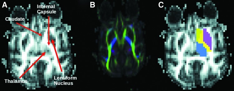
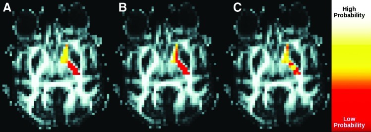
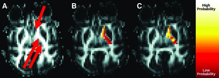
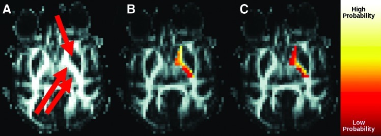
Understanding anatomical connectivity is crucial for improving outcomes of deep brain stimulation surgery. Tractography is a promising method for noninvasively investigating anatomical connectivity, but connections between subcortical regions have not been closely examined by this method. As many connections to subcortical regions converge at the internal capsule (IC), we investigate the connectivity through the IC to three subcortical nuclei (caudate, lentiform nucleus, and thalamus) in six macaques. We show that a statistical correction for a known distance-related artifact in tractography results in large changes in connectivity patterns. Our results suggest that care should be taken in using tractography to assess anatomical connectivity between subcortical structures.
Figures
Similar articles
-
Functional network mirrored in the prefrontal cortex, caudate nucleus, and thalamus: high-resolution functional imaging and structural connectivity.J Neurosci. 2014 Jul 9;34(28):9202-12. doi: 10.1523/JNEUROSCI.0228-14.2014. J Neurosci. 2014. PMID: 25009254 Free PMC article.
-
Feasibility of prefronto-caudate pathway tractography using high resolution diffusion tensor tractography data at 3T.J Neurosci Methods. 2010 Aug 30;191(2):249-54. doi: 10.1016/j.jneumeth.2010.06.026. Epub 2010 Jun 30. J Neurosci Methods. 2010. PMID: 20600311 Free PMC article.
-
Fronto-striatal connections in the human brain: a probabilistic diffusion tractography study.Neurosci Lett. 2007 May 29;419(2):113-8. doi: 10.1016/j.neulet.2007.04.049. Epub 2007 May 4. Neurosci Lett. 2007. PMID: 17485168 Free PMC article.
-
Deep brain stimulation: anatomical, physiological, and computational mechanisms.Network. 2011;22(1-4):186-207. doi: 10.3109/0954898X.2011.638356. Network. 2011. PMID: 22149679 Review. No abstract available.
-
Tractography: where do we go from here?Brain Connect. 2011;1(3):169-83. doi: 10.1089/brain.2011.0033. Epub 2011 Aug 30. Brain Connect. 2011. PMID: 22433046 Free PMC article. Review.
Cited by
-
Clinical Evaluation of Cingulum Bundle Connectivity for Neurosurgical Hypothesis Development.Neurosurgery. 2020 May 1;86(5):724-735. doi: 10.1093/neuros/nyz225. Neurosurgery. 2020. PMID: 31264700 Free PMC article.
-
Increased insula-putamen connectivity in X-linked dystonia-parkinsonism.Neuroimage Clin. 2017 Oct 28;17:835-846. doi: 10.1016/j.nicl.2017.10.025. eCollection 2018. Neuroimage Clin. 2017. PMID: 29527488 Free PMC article.
-
A Symmetry-Based Method to Infer Structural Brain Networks from Probabilistic Tractography Data.Front Neuroinform. 2016 Nov 4;10:46. doi: 10.3389/fninf.2016.00046. eCollection 2016. Front Neuroinform. 2016. PMID: 27867354 Free PMC article.
-
Automatic target validation based on neuroscientific literature mining for tractography.Front Neuroanat. 2015 May 27;9:66. doi: 10.3389/fnana.2015.00066. eCollection 2015. Front Neuroanat. 2015. PMID: 26074781 Free PMC article.
-
Low-Intensity Focused Ultrasound Neuromodulation for Stroke Recovery: A Novel Deep Brain Stimulation Approach for Neurorehabilitation?IEEE Open J Eng Med Biol. 2023 Apr 5;4:300-318. doi: 10.1109/OJEMB.2023.3263690. eCollection 2023. IEEE Open J Eng Med Biol. 2023. PMID: 38196977 Free PMC article.
References
-
- Basser PJ. Mattiello J. Lebihan D. Estimation of the effective self-diffusion tensor from the NMR spin echo. J Magn Reson B. 1994;103:247–254. - PubMed
-
- Basser PJ. Pierpaoli C. Microstructural and physiological features of tissues elucidated by quantitative-diffusion-tensor MRI. J Magn Reson B. 1996;111:209–219. - PubMed
-
- Behrens TEJ, et al. Non-invasive mapping of connections between human thalamus and cortex using diffusion imaging. Nat Neurosci. 2003;6:750–757. - PubMed
-
- Coenen VA. Mädler B. Schiffbauer H. Urbach H. Allert N. Individual fiber anatomy of the subthalamic region revealed with DTI—a concept to identify the DBS target for tremor suppression. Neurosurgery. 2011;68:1069–1076. - PubMed
Publication types
MeSH terms
Grants and funding
LinkOut - more resources
Full Text Sources
Research Materials
