

Diagnosis and treatment in neuro-oncology: an oncological perspective
- PMID: 22433832
- PMCID: PMC3473904
- DOI: 10.1259/bjr/18061999
Diagnosis and treatment in neuro-oncology: an oncological perspective
Abstract
Although brain tumours are rare compared with other malignancies, they are responsible, in many cases, for severe physical and cognitive disability and have a high case fatality rate (13% overall survival at 5 years). Gliomas account for over 60% of primary brain tumours and usually present with one or more symptoms of raised intracranial pressure, progressive neurological deficit, seizures, focal or global cognitive decline. The diagnosis is made by a combination of imaging and histological examination of tumour specimen. Contrast-enhanced MRI is the gold standard imaging modality and provides highly sensitive anatomical information about the tumour. Advanced imaging modalities provide complementary information about brain tumour metabolism, blood flow and ultrastructure and are being increasingly incorporated into routine clinical sequences. Imaging is essential for guiding surgery and radiotherapy treatments and for monitoring response to, and progression of, therapy. However, changes in imaging over time may be misinterpreted and lead to incorrect assumptions about the effectiveness of treatments. Thus, the disappearance of contrast enhancement and resolution of oedema after anti-angiogenesis treatments is seen early while conventional T(2) weighted/FLAIR sequences demonstrate continual tumour growth (pseudoregression). Conversely imaging may suggest lack of efficacy of treatment e.g. increasing tumour size and contrast enhancement following chemoradiation for malignant gliomas (pseudoprogression), which then stabilise or resolve after a few months of continued treatment and that paradoxically may be associated with a better outcome. These factors have led to a re-evaluation of the role of standard sequences in the assessment of treatment response spurning interest in the development of quantitative biomarkers.
Figures
References
-
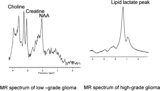
- Leclerc X, Huisman AGM, Sorensen AG. The potential of proton magnetic resonance spectroscopy (1H-MRS) in the diagnosis and management of patients with brain tumors. Curr Opinion Oncol 2002;14:292–8 - PubMed
Publication types
MeSH terms
Substances
LinkOut - more resources
Full Text Sources
Medical
