. 2012 May 11;287(20):16467-76.
doi: 10.1074/jbc.M112.349274. Epub 2012 Mar 20.

Interactions of nucleolin and ribosomal protein L26 (RPL26) in translational control of human p53 mRNA

Affiliations

Interactions of nucleolin and ribosomal protein L26 (RPL26) in translational control of human p53 mRNA

Jing Chen et al. J Biol Chem. .

Abstract

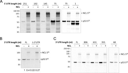
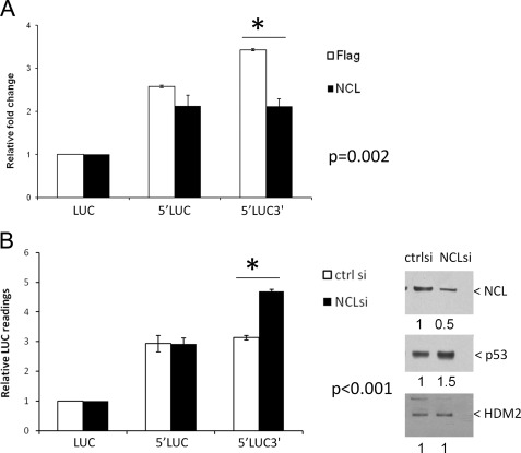
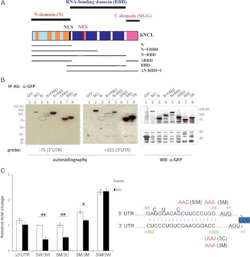
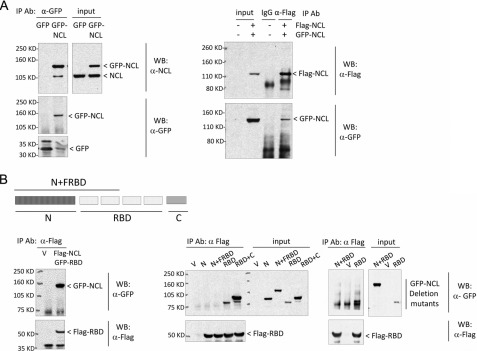
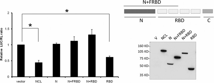
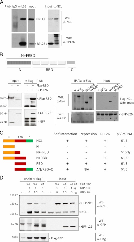
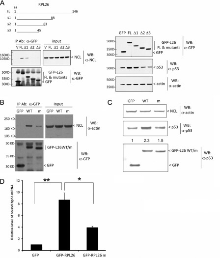
Ribosomal protein RPL26 enhances p53 translation after DNA damage, and this regulation depends upon interactions between the 5'- and 3'-UTRs of human p53 mRNA (Takagi, M., Absalon, M. J., McLure, K. G., and Kastan, M. B. (2005) Cell 123, 49-63; Chen, J., and Kastan, M. B. (2010) Genes Dev. 24, 2146-2156). In contrast, nucleolin (NCL) suppresses the translation of p53 mRNA and its induction after DNA damage. We confirmed reports that RPL26 and NCL interact with each other and then explored the potential role of this interaction in the translational control of p53 after stress. NCL repression of p53 translation utilizes both the 5'- and 3'-UTRs of p53 mRNA, and NCL binds to the same 5'-3'-UTR interaction region that is critical for the recruitment of RPL26 to p53 mRNA after DNA damage. We also found that NCL is able to oligomerize, consistent with a model in which NCL stabilizes this double-stranded RNA structure. We found that the RNA-binding domain of NCL participates in binding to p53 mRNA, is required for both NCL dimerization and NCL-mediated translational repression, and is the domain of NCL that interacts with RPL26. Excessive RPL26 disrupts NCL dimerization, and point mutations in the NCL-interacting region of RPL26 reduce NCL-RPL26 interactions and attenuate both RPL26 binding to human p53 mRNA and p53 induction by RPL26. These observations suggest a model in which the base pairings in the p53 UTR interaction regions are critical for both translational repression and stress induction of p53 by NCL and RPL26, respectively, and that disruption of a NCL-NCL homodimer by RPL26 may be the switch between translational repression and activation after stress.


Figures

FIGURE 1.
FIGURE 1.
Both 5′- and 3′-UTR sequences of human p53 mRNA are required for translational repression by NCL. A, the 5′-UTR of human p53 mRNA is required for translational repression of p53 by NCL in vitro. NCL repression of translation was evaluated in rabbit reticulocyte lysates containing p53 cDNAs with variable lengths of 5′-UTR sequences (as indicated) plus the full-length p53 coding sequence and the 3′-UTR without (−) or with (+) the NCL coding sequence added. The expression of newly synthesized, [35S]methionine-labeled protein was assessed by autoradiography. The panels merge the results of two independent experiments with different exposure times of the film. B, the 3′-UTR of human p53 mRNA is required for translational repression by NCL in vitro. Translation of p53 mRNA containing 75 bases of the 5′-UTR and the p53 coding sequence without (Δ3′UTR) or with (FL) the full-length 3′-UTR was evaluated in rabbit reticulocyte lysates without (−) or with (+) the NCL coding sequence. The expression of newly synthesized, [35S]methionine-labeled protein and total p53 protein was assessed by autoradiography. The band intensity of 35S-labeled p53 protein in each lane was quantitated using NIH ImageJ software and compared with that in the first lane, and the -fold changes are shown under the panel. C, the first 355 nucleotides (nt) of the p53 3′-UTR are required for nucleolin repression in vitro. p53 cDNA (75 nucleotide of the 5′-UTR plus the full-length coding sequence) with variable lengths of the 3′-UTR sequence was coexpressed without (−) or with (+) the NCL coding sequence in rabbit reticulocyte lysates. The expression of newly synthesized, [35S]methionine-labeled protein was assessed by autoradiography.
FIGURE 2.
FIGURE 2.
Both 5′- and 3′-UTR sequences of human p53 mRNA are required for translational repression by NCL in cells. A, overexpression of NCL represses the translation of human p53 mRNA in the presence of both 5′- and 3′-UTR sequences. MCF-7 stable cell lines were established to constitutively express a firefly luciferase reporter gene without any UTR sequences of human p53 mRNA (LUC) or containing the 145-nucleotide p53 5′-UTR with (5′LUC3′) or without (5′LUC) the full-length p53 3′-UTR. FLAG-tagged NCL (NCL) or empty vector (Flag) was cotransfected with a Renilla luciferase (19) expression vector (internal control; RL) into these stable cell lines. 24 h post-transfection, modulation of the reporter gene expression was assessed by comparing the LUC/RL ratio of each samples with that of LUC samples. The error bars represent the mean ± S.D. for three independent experiments. *, the p value was calculated using Student's t test. B, knock down of NCL in cells enhances the translation of human p53 mRNA in the presence of both 5′- and 3′-UTR sequences. 40 μm NCL siRNA duplex (NCLsi) (1) or non-target control siRNA (ctrl si) was introduced into the above MCF-7 stable reporter cell lines. 3 days post-transfection, the endogenous NCL levels was assessed by immunoblotting (right panels). Modulation of the reporter gene expression was assessed by comparing the firefly luciferase reading of each sample with that of LUC samples (left panel). The error bars represent the mean ± S.D. for three independent experiments. *, the p value was calculated using Student's t test. The band intensity of NCL (upper right panel), p53 (middle right panel) and HDM2 (lower right panel) proteins was quantitated using NIH ImageJ software, and -fold changes in the intensity compared with those in the NCL siRNA duplex sample are shown under the panels.
FIGURE 3.
FIGURE 3.
NCL repression requires base pairings within UTR interaction region. A, schematic diagram of nucleolin functional domains adopted from Ref. and nucleolin deletion mutants generated in this study. NLS, nuclear localization signal; NES, nuclear export signal; hNCL, human NCL; FRBD, first half of the RBD. B, NCL and its RBD directly and independently bind both the 5′- and 3′-UTRs of human p53 mRNA. GFP-tagged NCL deletion mutants were transiently transfected into H1299 cells and immunoprecipitated (IP) with anti-GFP antibody. The immunoprecipitated proteins were hybridized with a 32P-5′-end-labeled 5′-UTR (−75 to +1; left panel) or 3′-UTR (first 355 nucleotides; middle panel) RNA probe for Northwestern blot analysis. The immunoprecipitated proteins were also detected by Western blot (WB) analysis using anti-GFP antibody (right panel). C, disruption of the base pairings in UTR interaction region blunts NCL repression. Mutations were introduced into the last 3 bp of the 5′- and 3′-UTR interaction regions (5M/3M; depicted in the diagram on the right-hand side with mutations labeled in red), the middle 3 bases of the 5′-UTR in this region (5IM/3W), or a compensatory mutation (5M/3C) (2) that restores complementarity of the last 3 bp. The Dual-Luciferase reporter assay was performed as described under “Experimental Procedures.” Data shown are the mean ± S.D. for three independent experiments. p values were calculated using Student's t test. *, p < 0.01; **, p < 0.001.
FIGURE 4.
FIGURE 4.
NCL complexes with itself at RBD. A, NCL interacts with itself. GFP-tagged NCL was transfected (left panels) or cotransfected with FLAG-tagged NCL (right panels) into MCF-7 cells. GFP- or FLAG-tagged NCL was immunoprecipitated (IP) by anti-GFP or anti-FLAG antibody, respectively, and the bound protein was assessed by immunoblot analysis using anti-nucleolin (upper left panels) and anti-GFP (lower left panels) or anti-FLAG (upper right panel) and GFP (lower right panel) antibodies. 1–2 mg of total cell lysate was used for co-immunoprecipitation in each sample, and 10 μg of lysate was used as input. WB, Western blot. B, NCL interacts with itself at the RBD. The FLAG-tagged NCL RBD was cotransfected with GFP-NCL (left panels); with the GFP-tagged NCL N-terminal domain (N), the N-terminal domain plus the first half of the RBD (N+FRBD), the RBD, or the RBD plus the C-terminal domain (RBD+C) (middle panels); or with the N-terminal domain plus the full-length RBD (N+RBD) (right panels) into MCF-7 cells. FLAG-RBD was immunoprecipitated from cells using anti-FLAG antibody, and the bound proteins were detected by anti-FLAG (lower panels) or anti-GFP (upper panels) antibody. In these experiments, 3 mg of total cell lysate was used for co-immunoprecipitation in each sample, and 30 μg of lysate was used as input. V, empty vector.
FIGURE 5.
FIGURE 5.
NCL self-interaction is required for translational repression. MCF-7 cells were transiently transfected with empty vector (V), FLAG-NCL, or FLAG-tagged NCL deletion mutants together with a firefly luciferase construct (LUC) containing a 145-nucleotide p53 5′-UTR and the full-length p53 3′-UTR plus a control Renilla luciferase expression construct (RL). A simple diagram of the deletion mutants used is included (upper right panel). The relative LUC/RL ratio was calculated by normalizing the LUC/RL ratio of each sample to the ratio in cells transfected with the empty vector and both luciferase reporters. Data shown are the mean ± S.D. for three independent experiments. *, p < 0.0001 (Student's t test) (left panel). The expression level of FLAG-tagged proteins was assessed by immunoblotting with anti-FLAG antibody (lower right panel). FRBD, first half of the RBD.
FIGURE 6.
FIGURE 6.
NCL interacts with RPL26. A, NCL interacts with RPL26 in cells. Endogenous NCL or RPL26 (L26) was immunoprecipitated (IP) with anti-nucleolin or anti-RPL26 antibody. The bound protein was assessed using appropriate antibodies. 1–2 mg of total cell lysate was used for co-immunoprecipitation in each sample, and 20 μg of lysate was used as input. WB, Western blot. B, the RBD of NCL is sufficient for NCL-RPL26 interaction. FLAG-tagged RBD (left panels) or FLAG-tagged NCL deletion mutants (del muts; right panels) were cotransfected with GFP-RPL26 into MCF-7 cells. FLAG-tagged proteins were immunoprecipitated with anti-FLAG antibody. The bound proteins were detected by anti-GFP antibody. 1–3 mg of total cell lysate was used for co-immunoprecipitation in each sample, and 30 μg of lysate was used as input. FRBD, first half of the RBD; V, empty vector. C, the schematic diagram summarizes the results from domain mapping experiments described in Figs. 3–6. D, RPL26 disrupts NCL self-interaction. The indicated amounts of FLAG-RBD, GFP-RPL26, and GFP-NCL were cotransfected into MCF-7 cells. FLAG-RBD was immunoprecipitated by anti-FLAG antibody, and the bound proteins were detected using the indicated antibodies. 6 mg of total cell lysate was used for co-immunoprecipitation in each sample, and 30 μg of lysate was used as input.
FIGURE 7.
FIGURE 7.
NCL-RPL26 interaction affects human p53 induction by RPL26. A, a deletion mutant of RPL26 lost RPL26 binding to NCL (left panels) and the ability of RPL26 to induce p53 in cells (right panels). GFP-tagged RPL26 deletion mutants were transiently transfected into MCF-7 cells and immunoprecipitated (IP) using anti-GFP antibody. The bound NCL was detected using anti-nucleolin antibody (left panels). In this co-immunoprecipitation experiment, 30% of total cell lysate was used as input. The basal p53 protein level was also assessed in these samples before immunoprecipitation by immunoblotting (right panels). aa, amino acids; FL, full-length; V, empty vector; WB, Western blot. B, a point mutant of RPL26 lost RPL26 binding to NCL. GFP-tagged RPL26 point mutants (m; nine alanine replacements of amino acids 80–88) were transiently transfected into MCF-7 cells and immunoprecipitated using anti-GFP antibody 24 h post-transfection. The bound NCL was detected using anti-nucleolin antibody. 3 mg of total cell lysate was used for co-immunoprecipitation in each sample, and 20 μg of lysate was used as input. C, a point mutant of RPL26 lost RPL26 ability to induce p53 in cells. GFP-tagged RPL26 point mutants were transiently transfected into MCF-7 cells, and 24 h post-transfection, the basal p53 protein level was assessed by immunoblotting. D, a point mutant of RPL26 lost RPL26 binding to p53 mRNA in cells. GFP-tagged RPL26 point mutants were transiently transfected into MCF-7 cells and immunoprecipitated using anti-GFP antibody 24 h post-transfection. The bound p53 mRNA was measured by real-time RT-PCR. The bar graph shows the ratio of the bound p53 mRNA level compared with that seen in cells expressing GFP protein. The error bars represent the mean ± S.D. for three experiments. p values were calculated using Student's t test. *, p < 0.01; **, p < 0.001.

References

    1. Takagi M., Absalon M. J., McLure K. G., Kastan M. B. (2005) Regulation of p53 translation and induction after DNA damage by ribosomal protein L26 and nucleolin. Cell 123, 49–63 - PubMed
    1. Chen J., Kastan M. B. (2010) 5′-3′-UTR interactions regulate p53 mRNA translation and provide a target for modulating p53 induction after DNA damage. Genes Dev. 24, 2146–2156 - PMC - PubMed
    1. Kastan M. B., Bartek J. (2004) Cell cycle checkpoints and cancer. Nature 432, 316–323 - PubMed
    1. Levine A. J., Oren M. (2009) The first 30 years of p53: growing ever more complex. Nat. Rev. Cancer 9, 749–758 - PMC - PubMed
    1. Ashcroft M., Vousden K. H. (1999) Regulation of p53 stability. Oncogene 18, 7637–7643 - PubMed

Publication types

MeSH terms