Review
. 2012 Jul;32(7):1426-54.
doi: 10.1038/jcbfm.2012.20. Epub 2012 Mar 21.

The development, past achievements, and future directions of brain PET

Collaborators, Affiliations
Review

The development, past achievements, and future directions of brain PET

Terry Jones et al. J Cereb Blood Flow Metab. 2012 Jul.

Abstract

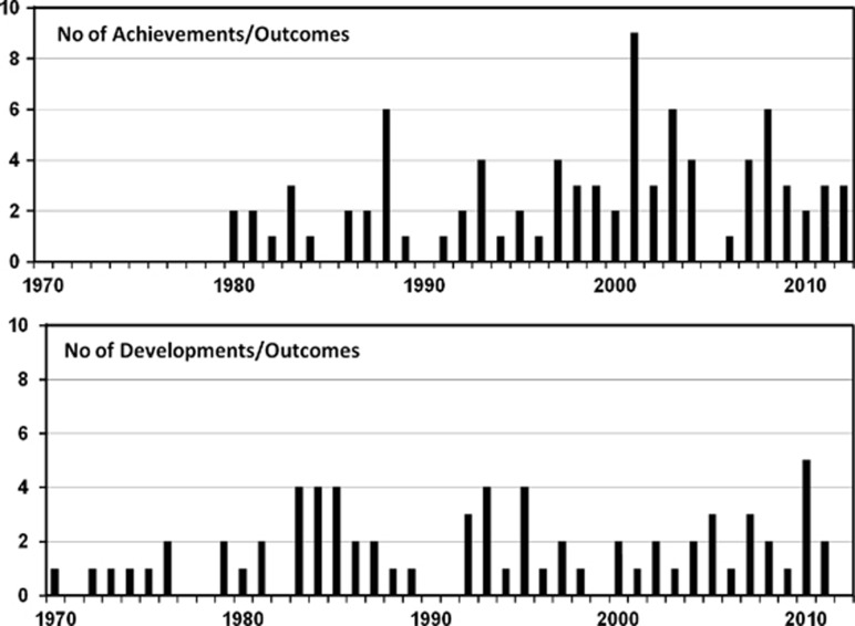
The early developments of brain positron emission tomography (PET), including the methodological advances that have driven progress, are outlined. The considerable past achievements of brain PET have been summarized in collaboration with contributing experts in specific clinical applications including cerebrovascular disease, movement disorders, dementia, epilepsy, schizophrenia, addiction, depression and anxiety, brain tumors, drug development, and the normal healthy brain. Despite a history of improving methodology and considerable achievements, brain PET research activity is not growing and appears to have diminished. Assessments of the reasons for decline are presented and strategies proposed for reinvigorating brain PET research. Central to this is widening the access to advanced PET procedures through the introduction of lower cost cyclotron and radiochemistry technologies. The support and expertize of the existing major PET centers, and the recruitment of new biologists, bio-mathematicians and chemists to the field would be important for such a revival. New future applications need to be identified, the scope of targets imaged broadened, and the developed expertize exploited in other areas of medical research. Such reinvigoration of the field would enable PET to continue making significant contributions to advance the understanding of the normal and diseased brain and support the development of advanced treatments.


Figures

Figure 1
Figure 1
(A) The first reported image of regional human brain metabolism in 1973 (Jones et al, 1976). This was recorded as a lateral view using the Massachusetts General Hospital positron camera (Burnham and Brownell, 1972) to image the steady-state distribution of radioactivity while continuously inhaling 15O2 as a tracer of oxygen utilization. Also shown is the steady-state distribution of radioactivity while continuously inhaling C15O to label red cells and hence delineate the cerebral vascular volume. (B) Anterior-posterior view of the steady distribution of radioactivity while continuously inhaling 15O2. The camera data have been focused on four equally spaced vertical planes.
Figure 2
Figure 2
Positron emission tomography (PET) images depicting cerebral blood flow (CBF), oxygen extraction ratio (OER), and cerebral metabolic rate of oxygen (CMRO2) of a patient after a left hemisphere transient ischemic attack (1ST), 7 hours after a major stroke (2ND), and 4 days after the stroke (3RD). The high oxygen extraction ratio seen within hours after the stroke fell in association with a decline in cortical oxygen metabolism (Wise et al, 1983).
Figure 3
Figure 3
Positron emission tomography (PET) images showing percentage reductions in regional glucose utilization and acetylcholinesterase (AChE) activity in Alzheimer's disease (AD) patients compared with controls, using [18F]-FDG and N-[11C] methylpiperidyl propionate, respectively. Glucose focal hypometabolism was focally prominent in posterior cingular gyrus and parietal cortex. Loss of AChE activity was even more diffuse and involved the entire neocortex, as well as the hippocampus (Kuhl et al, 1999). CMRglu, cerebral metabolic rate of glucose.
Figure 4
Figure 4
(A) [11C]raclopride positron emission tomography (PET) images through the caudate/putamen level of a schizophrenic patient treated with placebo (left) and haloperidol (right) as described in Farde et al (1992). (B) The suggested distinct thresholds for antipsychotic effects and extrapyramidal syndromes (EPSs) as induced by classical antipsychotic drugs. Owing to the hyperbolic relationship between occupancy of the D2 receptor and dose of antipsychotic drug (or plasma concentration) there is a rather narrow interval for optimal therapeutic treatment. Figure modified from Farde (1996).
Figure 5
Figure 5
(A) Straital [18F]16-fluoro-L-DOPA ([18F]F-DOPA) summation image. (B) Individual and group striatal [18F]F-DOPA Ki values (influx rate constants) for normal controls, subjects with At-Risk Mental Status (ARMS) and those with schizophrenia, showing significantly higher (P<0.05) Ki values in schizophrenia patients compared with controls. (C) Positive correlation between total Comprehensive Assessment of At-Risk Mental States (CAARMS) score (higher score indicates greater severity of prodromal symptoms) and Ki values (Howes et al, 2009).
Figure 6
Figure 6
Distribution of aerobic glycolysis in resting human brain using a glycolytic index (n=33, groupwise t test, ∣Z∣>4.4, P<0.0001, cluster>99, corrected for multiple comparisons). Specifically, regions with significantly high glycolysis include bilateral prefrontal cortex, bilateral lateral parietal lobe, posterior cingulate/precuneus, gyrus rectus, bilateral lateral temporal gyrus, and bilateral caudate nuclei. In contrast, cerebellum and bilateral inferior temporal gyrus have significantly low levels of aerobic glycolysis (Vaishnavi et al, 2010).
Figure 7
Figure 7
Chronology plots showing the significant ‘developmental and methodological' outcomes in brain PET and the reporting of ‘achievement' outcomes of new or consensus changing information on the human brain. Only one reference for a given outcome is scored. This analysis has not been based on a systematic review of the literature but on the references contained in this review. A large and important area of methodological developments is not directly represented, namely those that have occurred in radio synthetic chemistry, cyclotrons, and their production targetry.

References

    1. Abanades S, Van Der Aart J, Barletta JAR, Marzano C, Searle GE, Salinas CA, Ahmad JJ, Reiley RR, Pampols-Maso S, Zamuner S, Cunningham VJ, Rabiner EA, Laruelle MA, Gunn RN. Prediction of repeat-dose occupancy from single-dose data: characterisation of the relationship between plasma pharmacokinetics and brain target occupancy. J Cereb Blood Flow Metab. 2011;31:944–952. - PMC - PubMed
    1. Ackerman RH, Correia JA, Alpert NM, Baron JC, Gouliamos A, Grotta JC, Brownell GL, Taveras JM. Positron imaging in ischemic stroke disease using compounds labeled with oxygen 15. Initial results of clinicophysiologic correlations. Arch Neurol. 1981;38:537–543. - PubMed
    1. Alavi A, Reivich M. Guest editorial: the conception of FDG-PET imaging. Semin Nucl Med. 2002;32:2–5. - PubMed
    1. Albin RL, Minoshima S, D'Amato CJ, Frey KA, Kuhl DA, Sima AA. Fluoro-deoxyglucose positron emission tomography in diffuse Lewy body disease. Neurology. 1996;47:462–466. - PubMed
    1. Andersson JD, Pierson ME, Finnema SJ, Gulyas B, Heys R, Elmore CS, Farde L, Halldin C. Development of a PET radioligand for the central 5-HT1B receptor: radiosynthesis and characterization in cynomolgus monkeys of eight radiolabeled compounds. Nucl Med Biol. 2011;38:261–272. - PubMed

MeSH terms