. 2012 Aug;32(8):1484-95.
doi: 10.1038/jcbfm.2012.33. Epub 2012 Mar 21.

Investigating the metabolic changes due to visual stimulation using functional proton magnetic resonance spectroscopy at 7 T

Affiliations

Investigating the metabolic changes due to visual stimulation using functional proton magnetic resonance spectroscopy at 7 T

Yan Lin et al. J Cereb Blood Flow Metab. 2012 Aug.

Abstract

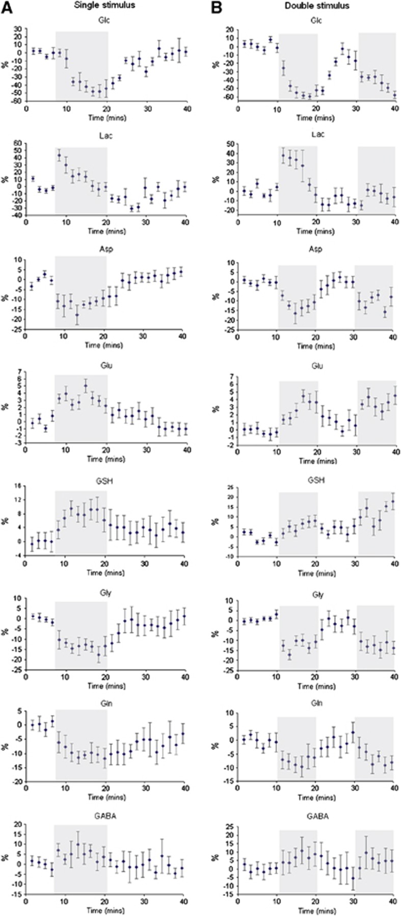
Proton magnetic resonance spectroscopy ((1)H-MRS) has been used to demonstrate metabolic changes in the visual cortex on visual stimulation. Small (2% to 11%) but significant stimulation induced increases in lactate, glutamate, and glutathione were observed along with decreases in aspartate, glutamine, and glycine, using (1)H-MRS at 7 T during single and repeated visual stimulation. In addition, decreases in glucose and increases in γ-aminobutyric acid (GABA) were seen but did not reach significance. Changes in glutamate and aspartate are indicative of increased activity of the malate-aspartate shuttle, which taken together with the opposite changes in glucose and lactate, reflect the expected increase in brain energy metabolism. These results are in agreement with those of Mangia et al. In addition, increases in glutamate and GABA coupled with the decrease in glutamine can be interpreted in terms of increased activity of the neurotransmitter cycles. An entirely new observation is the increase of glutathione during prolonged visual stimuli. The similarity of its time course to that of glutamate suggests that it may be a response to the increased release of glutamate or to the increased production of reactive oxygen species. Together, these observations constitute the most detailed analysis to date of functional changes in human brain metabolites.


Figures

Figure 1
Figure 1
(A) (i) One frame of the randomly varying visual stimulus presented to subjects. (ii) Representative example of blood oxygen level-dependent (BOLD) response with the voxel position for magnetic resonance spectroscopy (MRS) indicated by a white square (the yellow box shows the shim volume). (B) 1H-MRS spectra for the single and double stimulation paradigm using data from 17 subjects (group analysis) during (i) rest and (ii) stimulation. Difference spectrum before (iii) and after compensation for the BOLD effect (iv). Peaks ascribed to lactate (Lac) (1.33 p.p.m.), glutamate (Glu) (2.35/3.75 p.p.m.), and glutathione (GSH) (2.92/2.97/3.78 p.p.m.) are clearly evident in the compensated difference spectrum. The simulation, obtained by summation of the LCModel spectra of the metabolites for which significant changes were detected, clearly reproduces all the major features of the compensated difference spectrum. It provides convincing explanations as to why some resonances associated with a particular metabolite are visible, whereas others, for example, the GSH resonances at 2.54 and 2.16  p.p.m., are not. Asp, aspartate; GABA, γ-aminobutyric acid; Gln, glutamine; Gly, glycine; NAA, N-acetylaspartate; PCr, phosphocreatine; PE, phosphorylethanolamine. The color reproduction of this figure is available on the Journal of Cerebral Blood Flow and Metabolism journal online.
Figure 2
Figure 2
1H 7 T magnetic resonance spectroscopy (MRS) spectrum (STEAM (STimulated Echo Acquisition Mode) sequence: echo time/mixing time/repetition time (TE/TM/TR)=15/17/3,000 milliseconds, BW 4,000 Hz, 4,096 points, NSA 32) obtained from a 2 × 2 × 2 cm3 volume in the visual cortex (uppermost spectrum), LCModel fit (second spectrum from top) in the range 0.2 to 4.2 p.p.m. and residual (third spectrum from top). The LCModel basis components, macromolecule, and baseline contributions to the fit are shown in the lower traces. Asp, aspartate; GABA, γ-aminobutyric acid; Gln, glutamine; Glu, glutamate; Gly, glycine; GPC, glycerophosphorylcholine; GSH, glutathione; Lac, lactate; NAA, N-acetylaspartate; NAAG, N-acetylaspartylglutamate; PCr, phosphocreatine; PE, phosphorylethanolamine; scyllo-Ins, scyllo-inositol.
Figure 3
Figure 3
(A) LCModel quantification of metabolite levels for the single visual stimulation paradigm during rest (6.6 minutes), stimulation (13.2 minutes), and recovery (19.8 minutes). (B) For the metabolites showing significant change on stimulation, the data have been further analyzed in blocks of 6.6 minutes duration: rest, stimulation, recovery, 1, 2, and 3 blocks, respectively. All data (mean±s.d.) are shown for metabolites for which the CRLB were lower than specified limits (N=9 for all metabolites except N (alanine (Ala))=5, N (sI, lactate (Lac), N-acetylaspartylglutamate (NAAG), glucose (Glc))=6, N (Phosphorylethanolamine (PE))=7). Asp, aspartate; GABA, γ-aminobutyric acid; Gln, glutamine; Glu, glutamate; Gly, glycine; GSH, glutathione; NAA, N-acetylaspartate; PCr, phosphocreatine; scyllo-Ins, scyllo-inositol; tCr, total Cr.
Figure 4
Figure 4
(A) LCModel quantification of metabolite levels for the repeated (double) visual stimulation paradigm, comparing rest (two 9.9-minute periods) and stimulation (two 9.9-minute periods). (B) Data for individual rest and stimulation blocks for metabolites demonstrating significant change on activation. All data (mean±s.d.) are shown for metabolites for which the CRLB were lower than specified limits (N=8 for all metabolites except N (alanine (Ala))=5, N (sI, phosphorylethanolamine (PE))=7, N (lactate (Lac))=6, N (glucose (Glc))=4). Asp, aspartate; GABA, γ-aminobutyric acid; Gln, glutamine; Glu, glutamate; Gly, glycine; GSH, glutathione; NAA, N-acetylaspartate; NAAG, N-acetylaspartylglutamate; PCr, phosphocreatine; scyllo-Ins, scyllo-inositol; tCr, total Cr.
Figure 5
Figure 5
Time courses of metabolites during the single (A) and double (B) visual stimulation paradigms. Concentration changes are expressed as a percentage relative to the average concentration during the first rest period, temporal resolution is 99 seconds. Data are mean±s.e.m. (with the number of subjects included in each time course the same as in Figures 3 and 4 for the single and double stimulus, respectively). The shaded areas show the periods of visual stimulation. Asp, aspartate; GABA, γ-aminobutyric acid; Glc, glucose; Gln, glutamine; Glu, glutamate; Gly, glycine; GSH, glutathione; Lac, lactate.

References

    1. Badar-Goffer RS, Bachelard HS, Morris PG. Cerebral metabolism of acetate and glucose studied by C-13-NMR spectroscopy. A technique for investigating metabolic compartmentation in the brain. Biochem J. 1990;266:133–139. - PMC - PubMed
    1. Boer J, Postema F, Plijter-Groendijk H, Korf J. Continuous monitoring of extracellular lactate concentration by microdialysis lactography for the study of rat muscle metabolism in vivo. Pflügers Archiv Eur J Physiol. 1991;419:1–6. - PubMed
    1. Boucard CC, Mostert JP, Cornelissen FW, De Keyser J, Oudkerk M, Sijens PE. Visual stimulation, 1H MR spectroscopy and fMRI of the human visual pathways. Eur Radiol. 2005;15:47–52. - PubMed
    1. Cabanes E, Confort-Gouny S, Le Fur Y, Simond G, Cozzone PJ. Optimization of residual water signal removal by HLSVD on simulated short echo time proton MR spectra of the human brain. J Magn Reson. 2001;150:116–125. - PubMed
    1. Chen W, Zhu X-H, Gruetter R, Seaquist ER, Adriany G, Ugurbil K. Study of tricarboxylic acid cycle flux changes in human visual cortex during hemifield visual stimulation using 1H-{13C} MRS and fMRI. Magn Reson Med. 2001;45:349–355. - PubMed

Publication types

LinkOut - more resources