. 2012 Apr;135(Pt 4):1128-40.
doi: 10.1093/brain/aws040. Epub 2012 Mar 20.

Compensatory premotor activity during affective face processing in subclinical carriers of a single mutant Parkin allele

Affiliations

Compensatory premotor activity during affective face processing in subclinical carriers of a single mutant Parkin allele

Silke Anders et al. Brain. 2012 Apr.

Abstract

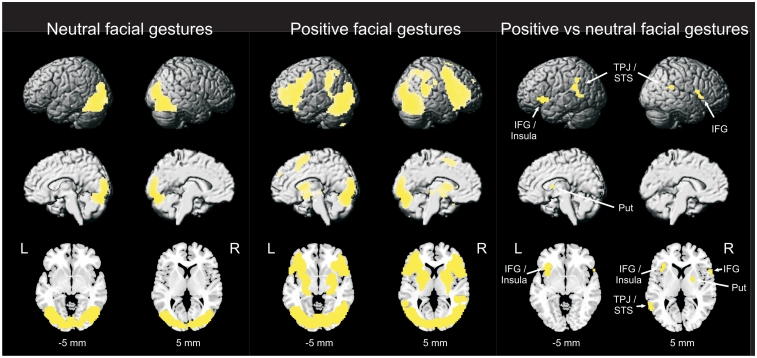
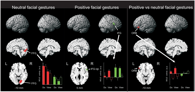
Patients with Parkinson's disease suffer from significant motor impairments and accompanying cognitive and affective dysfunction due to progressive disturbances of basal ganglia-cortical gating loops. Parkinson's disease has a long presymptomatic stage, which indicates a substantial capacity of the human brain to compensate for dopaminergic nerve degeneration before clinical manifestation of the disease. Neuroimaging studies provide evidence that increased motor-related cortical activity can compensate for progressive dopaminergic nerve degeneration in carriers of a single mutant Parkin or PINK1 gene, who show a mild but significant reduction of dopamine metabolism in the basal ganglia in the complete absence of clinical motor signs. However, it is currently unknown whether similar compensatory mechanisms are effective in non-motor basal ganglia-cortical gating loops. Here, we ask whether asymptomatic Parkin mutation carriers show altered patterns of brain activity during processing of facial gestures, and whether this might compensate for latent facial emotion recognition deficits. Current theories in social neuroscience assume that execution and perception of facial gestures are linked by a special class of visuomotor neurons ('mirror neurons') in the ventrolateral premotor cortex/pars opercularis of the inferior frontal gyrus (Brodmann area 44/6). We hypothesized that asymptomatic Parkin mutation carriers would show increased activity in this area during processing of affective facial gestures, replicating the compensatory motor effects that have previously been observed in these individuals. Additionally, Parkin mutation carriers might show altered activity in other basal ganglia-cortical gating loops. Eight asymptomatic heterozygous Parkin mutation carriers and eight matched controls underwent functional magnetic resonance imaging and a subsequent facial emotion recognition task. As predicted, Parkin mutation carriers showed significantly stronger activity in the right ventrolateral premotor cortex during execution and perception of affective facial gestures than healthy controls. Furthermore, Parkin mutation carriers showed a slightly reduced ability to recognize facial emotions that was least severe in individuals who showed the strongest increase of ventrolateral premotor activity. In addition, Parkin mutation carriers showed a significantly weaker than normal increase of activity in the left lateral orbitofrontal cortex (inferior frontal gyrus pars orbitalis, Brodmann area 47), which was unrelated to facial emotion recognition ability. These findings are consistent with the hypothesis that compensatory activity in the ventrolateral premotor cortex during processing of affective facial gestures can reduce impairments in facial emotion recognition in subclinical Parkin mutation carriers. A breakdown of this compensatory mechanism might lead to the impairment of facial expressivity and facial emotion recognition observed in manifest Parkinson's disease.


Figures

Figure 1
Figure 1
Design of the functional MRI study. The bar at the bottom represents one experimental run. Each participant underwent two runs. Colours indicate the six experimental conditions (green, do-smile; light green, view-smile; red, do-kiss; light red, view-kiss; blue, do-neutral; light blue, view-neutral). Note that ‘smile’ and ‘kiss’ conditions were fused in the analysis (see text).
Figure 2
Figure 2
Facial emotion recognition in controls and Parkin mutation carriers. (A) Average emotion recognition rates did not differ significantly between Parkin mutation carriers and controls; however, mutation carriers showed a significantly larger decrease in emotion recognition from clear-cut (90–10%) to fuzzy (70–30%) morphs. (B) There was no significant group × level × emotion interaction, indicating that the slight impairment of Parkin mutation carriers was not limited to any specific emotion. The asterisk indicates a significant effect (*P < 0.05); n.s = not significant. Parkin MC = Parkin mutation carriers.
Figure 3
Figure 3
Post-scan joy ratings. Parkin mutation carriers did not differ significantly from controls in self-reported joy in any condition.
Figure 4
Figure 4
Common BOLD activity during execution and observation of facial gestures in all participants. Statistical parametric maps for execution of facial gestures and for observation of facial gestures were individually thresholded at a false discovery rate of 0.05 (corrected for the whole volume) and inclusively masked such that only voxels are highlighted that show significant activity in both conditions. Only clusters that comprise at least 10 voxels in both contrasts are shown. IFG = inferior frontal gyrus; Put = putamen; STS = superior temporal sulcus; TPJ = temporoparietal junction.
Figure 5
Figure 5
Altered BOLD activity during processing of facial gestures in Parkin mutation carriers relative to controls. For visualization, conjoint statistical parametric maps for execution and observation of facial gestures are thresholded at a voxel-wise height threshold of P = 0.005, but only clusters are shown whose most significantly activated voxel does not exceed a false discovery rate of 0.05 (see text). The histogram at the bottom of each panel shows activity at the most significantly activated voxel in each map. Red bars = controls; green bars = Parkin mutation carriers; filled bars = positive facial expressions; open bars = neutral facial expressions. Error bars indicate standard error of the mean. FFG = fusiform gyrus; IFG op = inferior frontal gyrus, pars opercularis; ORB = inferior frontal gyrus, pars orbitalis.
Figure 6
Figure 6
Correlation of BOLD activity and facial emotion recognition ability/self-reported joy. (A and B) Overall correlation between BOLD activity and facial emotion recognition accuracy in all participants (group differences are removed from BOLD and behavioural data). (C and D) Slope between BOLD activity and facial emotion recognition accuracy in controls (red squares) and Parkin mutation carriers (green circles). Projections of the group means (indicated by the plus) on the x- and y-axes, respectively, indicate the group mean of BOLD/behavioural data. (E and F) Overall correlation between BOLD activity and post-scan joy ratings in all participants (group differences are removed from BOLD and behavioural data). (G and H) Slope between BOLD activity and post-scan joy ratings in controls (red squares) and Parkin mutation carriers (green circles). Projections of the group means (indicated by the plus) on the x- and y-axes, respectively, indicate the group mean of BOLD/behavioural data. X-axes represent the contrast of parameter estimates ‘positive-minus-neutral’, averaged across ‘do’ and ‘view’. Y-axes represent emotion recognition scores for fuzzy morphs (70–30%)/post-scan joy ratings (scale 1–7, ‘positive-minus-neutral’, see text), averaged across ‘do’ and ‘view’. Asterisks indicate a significant correlation (A, B, E and F) or a significant difference between slopes for controls and Parkin mutation carriers (C, D, G and H).

References

    1. Adolphs R, Damasio H, Tranel D, Cooper G, Damasio AR. A role for somatosensory cortices in the visual recognition of emotion as revealed by three-dimensional lesion mapping. J Neurosci. 2000;20:2683–90. - PMC - PubMed
    1. Alexander GE, Crutcher MD, DeLong MR. Basal ganglia-thalamocortical circuits: parallel substrates for motor, oculomotor, “prefrontal” and “limbic” functions. Prog Brain Res. 1990;85:119–46. - PubMed
    1. Alexander GE, DeLong MR, Strick PL. Parallel organization of functionally segregated circuits linking basal ganglia and cortex. Annu Rev Neurosci. 1986;9:357–81. - PubMed
    1. Andersson JL, Hutton C, Ashburner J, Turner R, Friston K. Modeling geometric deformations in EPI time series. Neuroimage. 2001;13:903–19. - PubMed
    1. Bastiaansen JA, Thioux M, Keysers C. Evidence for mirror systems in emotions. Philos Trans R Soc Lond B Biol Sci. 2009;364:2391–404. - PMC - PubMed

Publication types

MeSH terms