

Ankle reconstruction in type II fibular hemimelia
- PMID: 22434224
- PMCID: PMC3332325
- DOI: 10.1007/s11751-012-0129-4
Ankle reconstruction in type II fibular hemimelia
Abstract
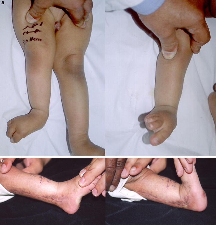
Ankle reconstruction prior to limb lengthening for was performed in 13 patients with fibular hemimelia with complete radiological absence of the fibula (type II). There were different degrees of absence of metatarsal rays. The hindfoot deformity was a heel valgus in 12 patients and equinovarus in 1 patient. The patients' ages ranged from 9 to 26 months. Excision of the fibular anlage was performed with lateral subtalar and ankle soft tissue releases to restore the ankle and subtalar joint relationships. In all cases, the fibular anlage ended distally in a cartilaginous lateral malleolar remnant that was fused to the talus in two patients. This fibular remnant was advanced distally and fixed to the tibia with 2 Kirschner wires to recreate an ankle mortise. The period of follow-up ranged from 12 to 38 months. All patients had a stable ankle without tendency to valgus deformity or subluxation. The ankle range of movement was a mean of 27.3° plantarflexion (25-30) and 18° dorsiflexion (15-20). Reconstruction of the ankle in type II fibular hemimelia using advancement of the cartilaginous lateral malleolar remnant has produced encouraging results in the short-term but longer follow-up is needed.
Figures

References
-
- Goh JC, Mech AMI, Lee EH. Biomechanical study on the load-bearing characteristics of the fibula and the effects of the fibular resection. Clin Orthop. 1992;279:223–228. - PubMed
-
- Lee EH, Goh JC, Helm R. Donor site morbidity following resection of the fibula. J Bone Joint Surg (Br) 1990;72-B:129–131. - PubMed
-
- Shogi H, Koshino T, Marcove RC. Subperiosteal resection of the distal portion of the fibula for aneurysmal bone cyst. J Bone Joint Surg (Am) 1970;52-A:1472–1476. - PubMed
-
- Achterman C, Kalamchi A. Congenital deficiency of the fibula. J Bone Joint Surg (Br) 1979;61-B:133–137. - PubMed
LinkOut - more resources
Full Text Sources
