. 2012 Oct 15;520(15):3528-52.
doi: 10.1002/cne.23104.

Biciliated ependymal cell proliferation contributes to spinal cord growth

Affiliations

Biciliated ependymal cell proliferation contributes to spinal cord growth

Clara Alfaro-Cervello et al. J Comp Neurol. .

Abstract

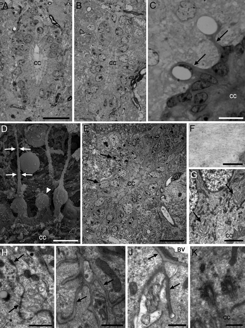
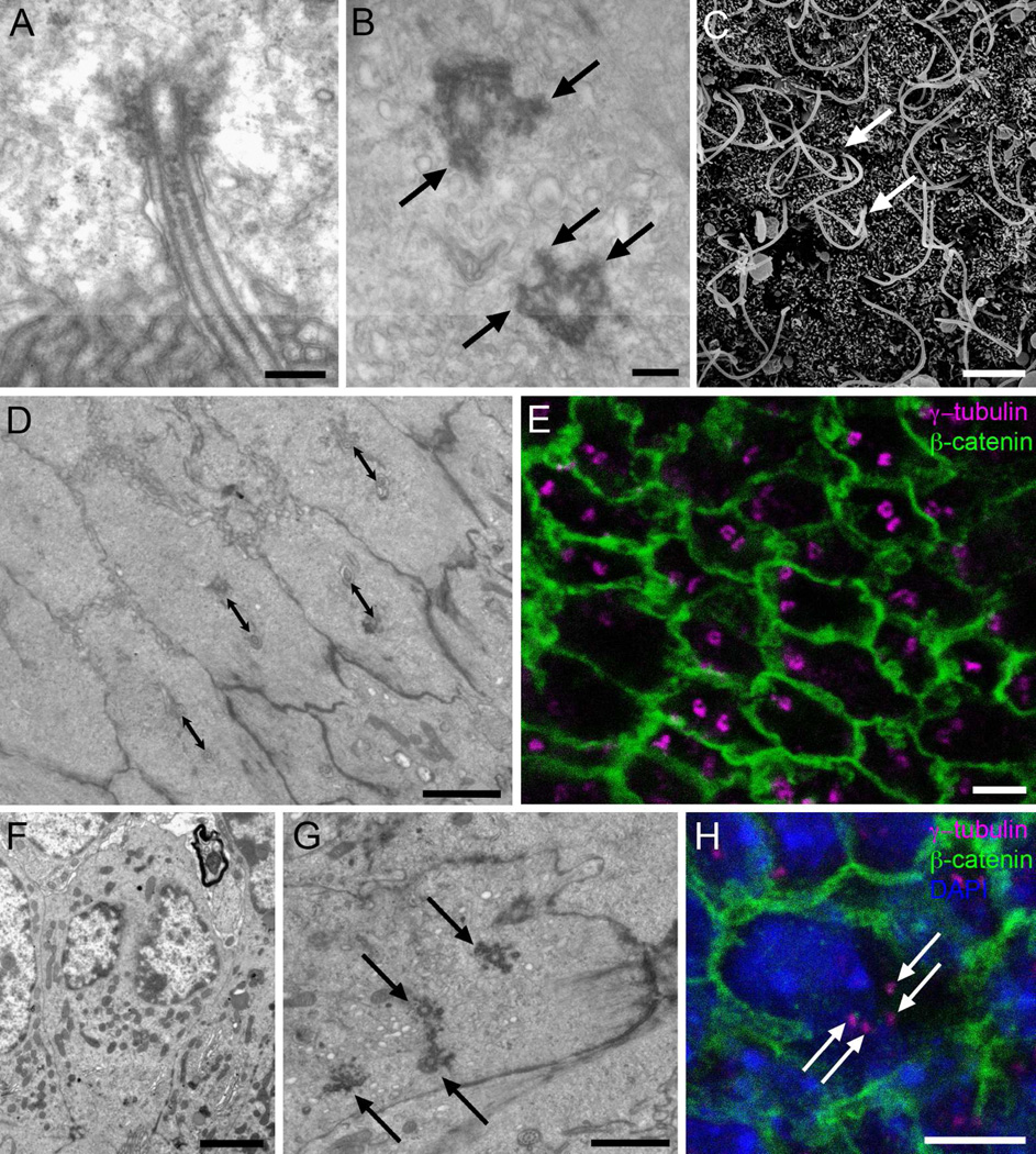
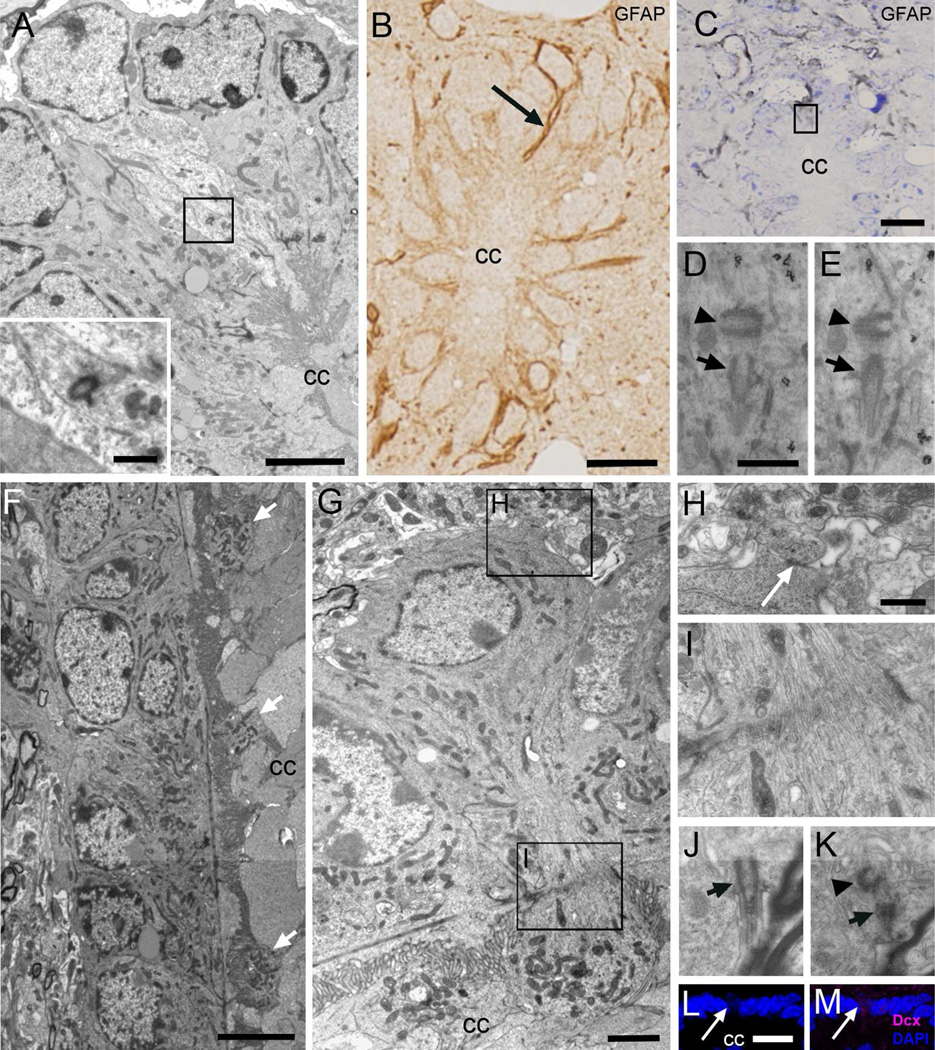
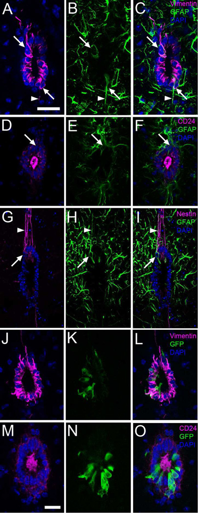
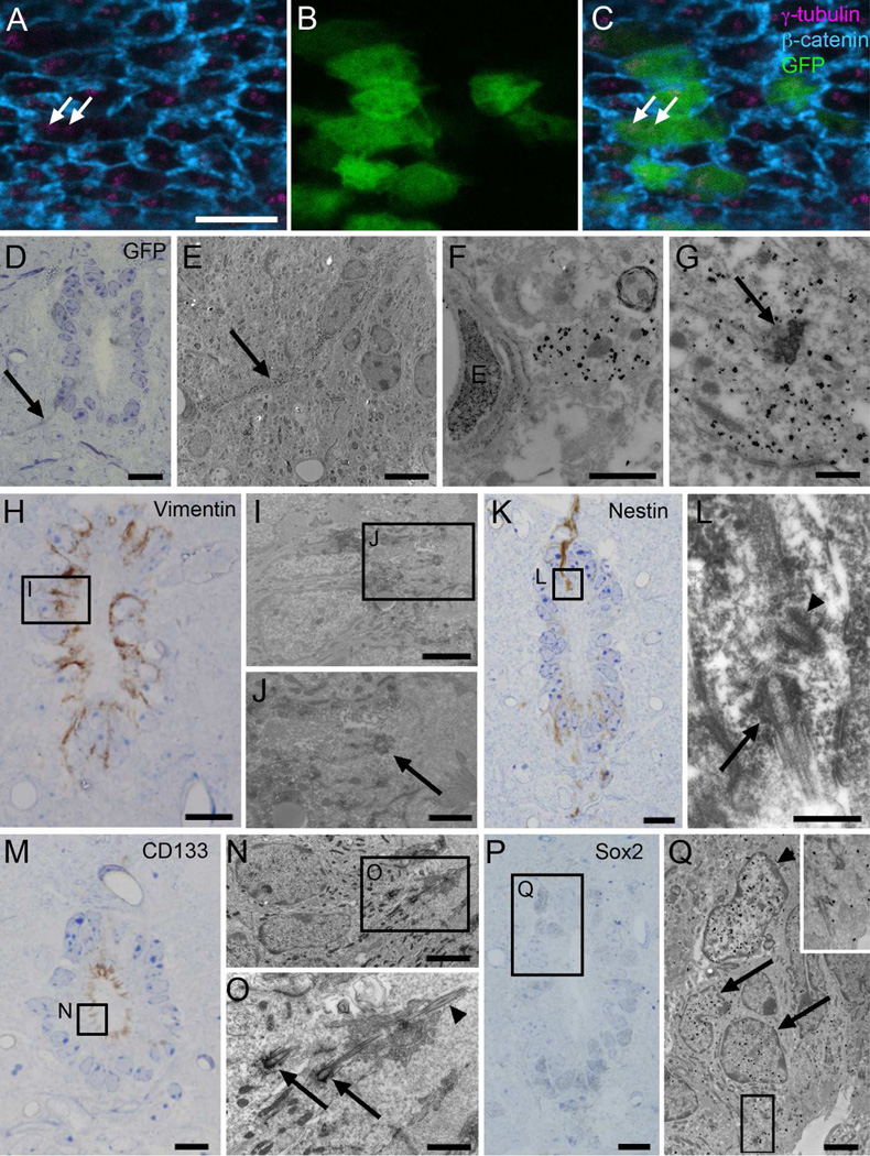
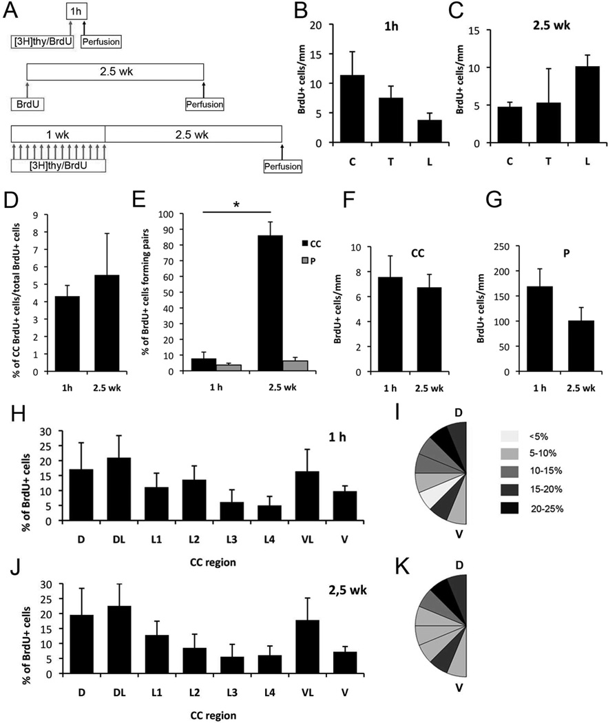
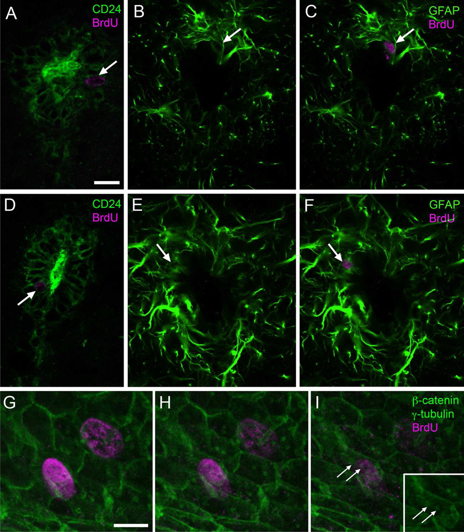
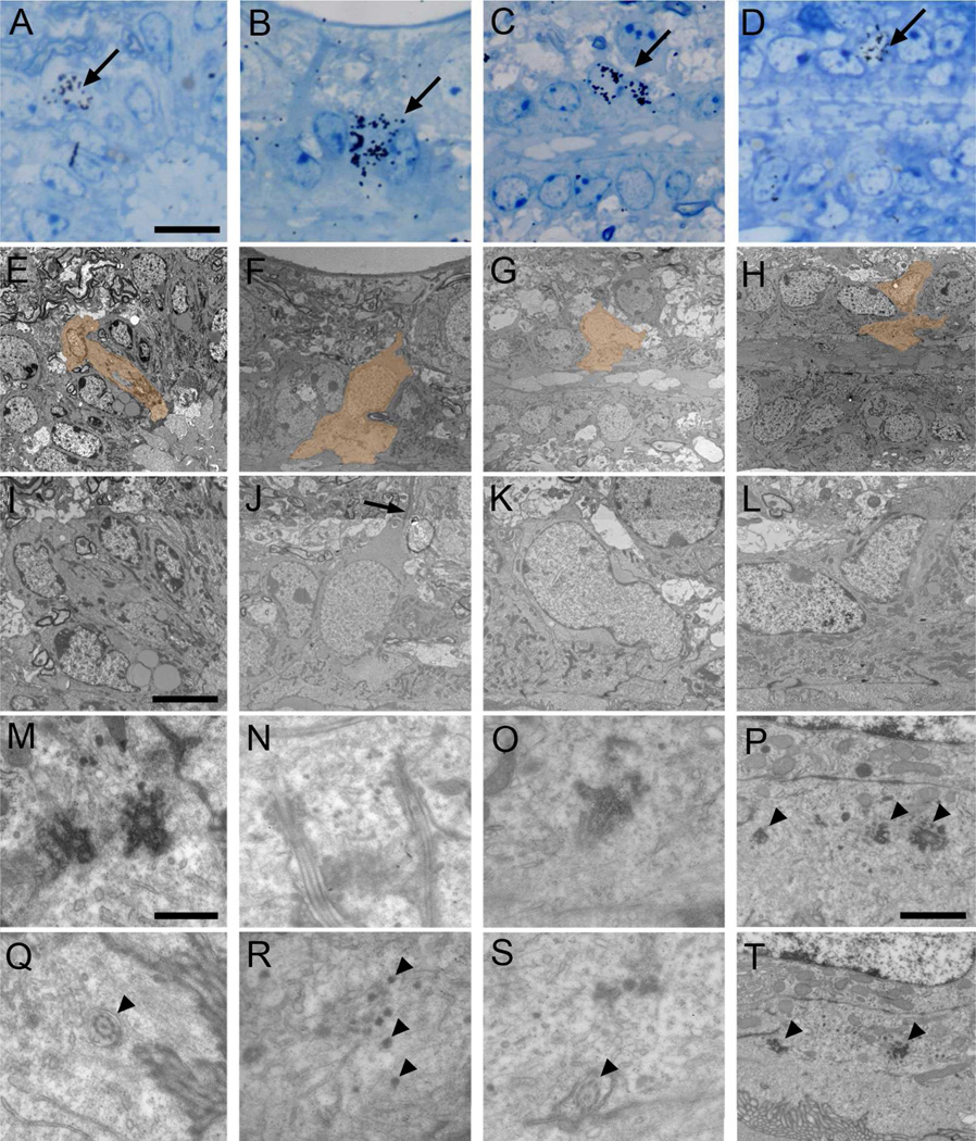
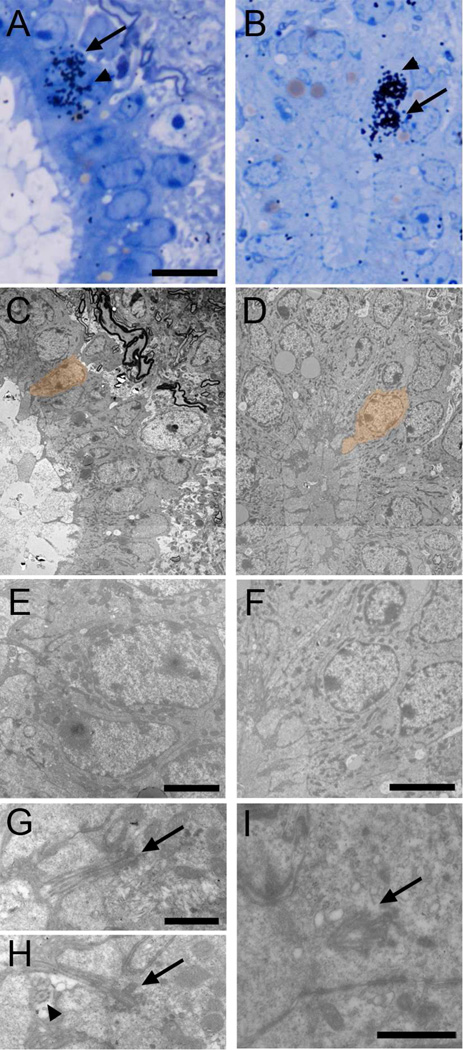
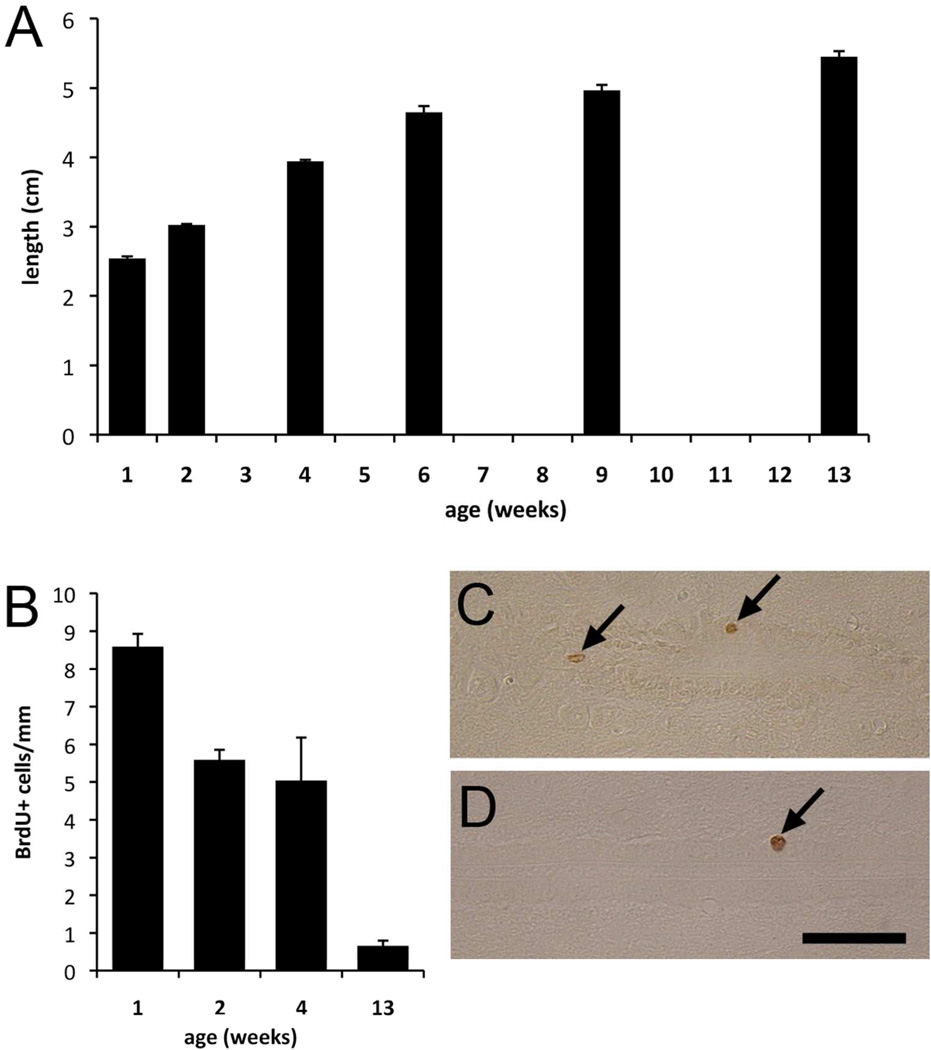
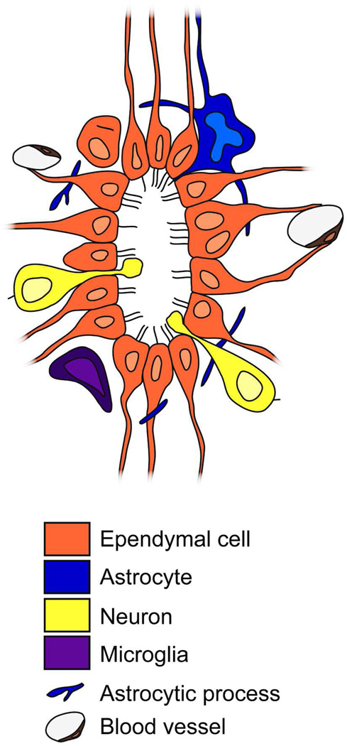
Two neurogenic regions have been described in the adult brain, the lateral ventricle subventricular zone and the dentate gyrus subgranular zone. It has been suggested that neural stem cells also line the central canal of the adult spinal cord. Using transmission and scanning electron microscopy and immunostaining, we describe here the organization and cell types of the central canal epithelium in adult mice. The identity of dividing cells was determined by 3D ultrastructural reconstructions of [(3) H]thymidine-labeled cells and confocal analysis of bromodeoxyuridine labeling. The most common cell type lining the central canal had two long motile (9+2) cilia and was vimentin+, CD24+, FoxJ1+, Sox2+, and CD133+, but nestin- and glial fibrillary acidic protein (GFAP)-. These biciliated ependymal cells of the central canal (Ecc) resembled E2 cells of the lateral ventricles, but their basal bodies were different from those of E2 or E1 cells. Interestingly, we frequently found Ecc cells with two nuclei and four cilia, suggesting they are formed by incomplete cytokinesis or cell fusion. GFAP+ astrocytes with a single cilium and an orthogonally oriented centriole were also observed. The majority of dividing cells corresponded to biciliated Ecc cells. Central canal proliferation was most common during the active period of spinal cord growth. Pairs of labeled Ecc cells were observed within the central canal in adult mice 2.5 weeks post labeling. Our work suggests that the vast majority of postnatal dividing cells in the central canal are Ecc cells and their proliferation is associated with the growth of the spinal cord.


Figures

Figure 1
Figure 1. Central canal ependymal (Ecc) cells
A) Toluidine blue-stained semithin section of the cervical spinal cord, where the central canal shows an oval shape. B) The lumbar central canal is dorsoventrally elongated. C) Central canal cells in the lateral wall, with radial expansions (arrows) in contact with blood vessels. D) Scanning electron microscopy (SEM) image of the canal surface (below). Some cells show long and thin basal processes (arrows), and other cells show rounder cell bodies, without radial process (arrowhead). E) Cells in the central canal are organized as a pseudostratified epithelium, with most nuclei located in the apical row, and some of them in basal position (arrows). The dorsal and ventral regions of the canal (up and below respectively) contain elongated Ecc cells with long radial processes. Blood vessels in close association to the canal can be observed. F) Detail of intermediate filaments of an Ecc cell. G) Horse-shoe shaped Golgi apparatuses polarized with the cis-side pointing at the apical surface (arrows). H) Large junction complexes are found apically between adjacent cells (arrows). I) Basal lamina network flowing through Ecc cells (arrows). J) Radial process of a central canal ependymal cell in contact with the basal lamina (arrows) of a blood vessel. K) Two large and electron-dense basal bodies of a biciliated Ecc cell. A–C are toluidine blue-stained semithin sections, and E–K are transmission electron microscopy (TEM) images. CC, central canal; BV, blood vessel. Scale bar in A (A–B) and E, 20 µm; C and D, 10 µm; F, 200nm; G, 2 µm; H, 1 µm; I, J and K, 500 nm.
Figure 2
Figure 2. Cilia and basal bodies of Ecc cells
A) Longitudinal TEM section of a cilium, showing the axoneme with a central pair of microtubules and a large electron-dense basal body. B) Tangential TEM section of the canal surface, showing basal bodies with multiple electron-dense appendages organized as radial spikes (arrows). C) SEM image of the canal surface, were long cilia can be observed as they emerge in parallel (arrows). No multiciliated cells were observed. D) Tangential TEM section of the canal surface, showing that biciliated Ecc cells´ basal bodies are oriented in the same direction and at a similar distance (double sided arrows: 1 µm). E) Immunofluorescence on central canal whole mount preparation. Donut-shaped basal bodies can be observed, stained with γ-tubulin (magenta), and limits between adjacent cells can be followed with β-catenin expression (green). F) TEM image showing an Ecc cell with two distinct nuclei, confirmed by serial ultrathin sectioning. G) En-face TEM section of the central canal surface showing one cell with four basal bodies (arrows). H) Confocal immunostaining on central canal whole mounts confirmed the presence of cells with two nuclei (DAPI, blue), and four basal bodies (γ-tubulin, magenta). Cell contours are labeled in green (β-catenin). Scale bar in A and B, 200 nm; C, 5 µm; D, E, F and H, 2 µm; G, 1 µm.
Figure 3
Figure 3. Astrocytes and neurons in contact with the central canal (Acc and Ncc cells)
A) Ultrastructure of an Acc cell with light cytoplasm in contact with the canal in the dorsal region in a transverse TEM section, showing an orthogonally oriented centriole (inset). B) Post-embedding immunocytochemistry on semithin sections, showing a thin GFAP+ expansion in contact with the canal lumen (arrow). C–E) Pre-embedding immunogold (detection of GFAP. C) Toluidine blue-stained semithin section showing grey immunogold-silver labeling for GFAP around the central canal and in thin expansions in contact with its lumen (boxed area). D–E) TEM serial sectioning of the box in C. One of the labeled cells displays a typical single cilium basal body (arrow) with an orthogonally oriented centriole (arrowhead). F) Longitudinal TEM section of the canal where globular expansions of several Ncc cells can be detected (arrows). G) These neurons show a round soma, and their cytoplasm contains abundant mitochondria and RER. H) Ncc cells establish axo-somatic synaptic contacts (arrow) in the surrounding neuropil. I) The apical narrowing contains abundant microtubules. J–K) Single cilium (arrow) with centriole (arrowhead) in a different Ncc cell that was serially reconstructed. L–M) Confocal immunofluorescence shows that Ncc cells express DCX (magenta) and their nuclei are weakly stained with DAPI (blue). CC, central canal. Scale bar in A and G, 2 µm, inbox, 500 nm; B, C and L, 10 µm; D (D–E) and H (H–K), 500 nm and; F, 5 µm.
Figure 4
Figure 4. Molecular markers in the central canal
A–C) Most central canal cells express vimentin intermediate filaments (magenta). Some cells also contain GFAP (green, arrows). GFAP is also frequent outside the canal. Some cells also express GFAP and vimentin in long processes (arrowheads). D–F) Nearly all canal cells express CD24 (magenta). Arrows point to a CD24- cell expressing GFAP (green). G–I) Nestin (magenta) was expressed predominantly in the dorsal and ventral regions of the central canal, especially in very long dorsal radial processes, which were frequently also positive for GFAP (green, arrowheads). The arrows point to a nestin+ cell also labeled with GFAP. J–O) Expression of FoxJ1 in the FoxJ1 ::Cre;Z/EG mice. J–L) FoxJ1 (green) is also expressed by vimentin+ cells (magenta). M–O) Immunofluorescence for GFP (green) and CD24 (magenta). Nuclei are stained with DAPI (blue). Scale bar in A (A–L), 20 µm; M (M–O), 10 µm.
Figure 5
Figure 5. Characterization of central canal cells
A–C) Confocal immunofluorescence on central canal whole mounts of the FoxJ1 ::Cre;Z/EG mice shows that GFP+ cells (green) display two basal bodies (γ-tubulin, magenta, arrows). β-catenin is shown in blue. D–G) Pre-embedding immunogold for GFP, showing several FoxJ1+ cells, one of them with a long radial process (arrows in D and E) in contact with a blood vessel. E) TEM of the same radial cell (arrow) labeled with silver-gold particles. F) Detail of the contact with the blood vessel’s basal lamina. G) Characteristic Ecc basal body (arrow) in a labeled cell. H–J) Pre-embedding staining for vimentin showed that labeled cells corresponded to Ecc cells with large electron-dense basal bodies (arrow). K–L) Nestin pre-embedding staining labeled cells with a single cilium (arrow) and centriole (arrowhead) in the dorsal and ventral regions of the canal. M–O) Pre-embedding staining for CD133 was detected along the apical surface of Ecc cells with large electron-dense basal bodies (arrows) and 9+2 axoneme (arrowhead). P–Q) Sox2 pre-embedding immunogold staining labeled both Acc cells (arrowhead) and Ecc cells (arrows) in the central canal. Inset shows characteristic basal bodies and cilia of Ecc cells. D, H, K, M and P are toluidine blue-stained semithin sections, which were re-embedded and serially cut for TEM analysis, as shown in E–G, I, J, L, N, O and Q. E, endothelial cell. Scale bar in A (A–C) and E, 5 µm; D, H, K, M and P, 10 µm; F, J, and O, 1 µm; G and L, 500 nm; I, N and Q, 2 µm.
Figure 6
Figure 6. Proliferation along the spinal cord
A) BrdU and [3H]thymidine injection protocols. B–C) Proliferation in the central canal and in the parenchyma does not significantly vary along cervical, thoracic and lumbar regions, 1 hour (1 h) as well as 2.5 weeks (2.5 wk) after BrdU injection. D) In the spinal cord, the percentage of labeled cells in the canal from the total number of spinal cord labeled cells did not vary between the two studied protocols. E) More than 80% of labeled cells after 2.5 weeks are forming pairs in the central canal. F–G) In the spinal cord central canal and parenchyma, no differences were observed between labeled cell numbers after 1 hour or 2.5 weeks. H–K) Distribution of BrdU+ cells along the dorsoventral axis in the central canal 1 hour (H–I) and 2.5 weeks (J–K) after BrdU injection. Labeled cells concentrated in dorsal and ventral regions, and fewer cells were found in the lateral canal walls. This distribution was very similar in both groups of study. A–G) C, cervical; T, thoracic; L, lumbar; CC, central canal; P, parenchyma. H–K) CC, central canal; D, dorsal; DL, dorsolateral; L1-L4, lateral; VL, ventrolateral; V, ventral. Error bars show SEM.
Figure 7
Figure 7. Identification of proliferating and generated cell types with immunofluorescence
A–F) Most BrdU+ cells (arrows, magenta) 1 hour (A–C) or 2.5 weeks (D–F) after injection express CD24 and occasionally GFAP (green). E) Whole mount staining showed BrdU+ cells (magenta) with two basal bodies (γ-tubulin, green, arrows). β-catenin is also shown in green. Scale bar in A (A–F), 10 µm; G (G–I), 2 µm.
Figure 8
Figure 8. Identification of proliferating cell types with TEM
Autoradiography on animals sacrificed 1 hour after [3H]thymidine injection. Labeled cells were identified on toluidine blue-stained semithin sections (A-D, arrows), serial ultrathin sectioned and analyzed under the TEM (E–T). Electronmicrographs of cells pointed with an arrow and depicted in orange are shown. A) Labeled Ecc cell (E and I), showing apically two large and electron-dense basal bodies (M). In a different section, the 9+2 microtubule structure of the cilium can be observed (Q, arrowhead). B) A different biciliated Ecc cell identified on a longitudinal section (F and J). It shows a radial expansion (J, arrow) and two cilia cut longitudinally (N). Other sections of the reconstruction showed dense particles close to the basal bodies (R, arrowheads). C) Uniciliated Ecc cell labeled on a longitudinal section (G and K). The cell showed apically a cilium with a large electron-dense basal body (O) and 9+2 microtubule structure (S, arrowhead). D) Frequently, labeled ependymal cells (H and L) contained apically basal body portions and electron-dense particles (P and T, arrowheads). Scale bar in A (A–H), 10 µm; I (I–L), 5 µm; M (M–O and Q–S), 500 nm; P (P and T), 1 µm.
Figure 9
Figure 9. Identification of generated cell types with TEM
Autoradiography on animals sacrificed 2.5 weeks after [3H]thymidine injections. Labeled cells were identified on toluidine blue-stained semithin sections (A-B, arrows), serial ultrathin sectioned and analyzed under the TEM (C–I). Pairs of labeled cells were frequently observed (arrows and arrowheads). Electronmicrographs of cells pointed with an arrow and depicted in orange are shown. A) Labeled Ecc cell (C and E) with two cilia, appearing on two different ultrathin sections (G–H). These cilia showed 9+2 structure (arrowhead) and large basal bodies (arrows). B) Labeled uniciliated Ecc cell (D and F) showing the beginning of the cilium with a large basal body (I, arrow). Scale bar in A (A–D), 10 µm; E, 2 µm; F, 5 µm; G (G–H) and I, 1 µm.
Figure 10
Figure 10. Spinal cord growth and proliferation
A) The spinal cord continues growing postnatally, but the length extension decreases progressively from 1 to 13 weeks. B) Central canal proliferation decreases with the animal´s age. C–D) Longitudinal section showing BrdU labeled cells (arrows) in mice aged 2 (C) and 13 (D) weeks. Scale bar, 50 µm. Error bars show SEM.
Figure 11
Figure 11. Model for the mouse central canal organization
Transverse schematic view showing the different cell types found in the central canal. We describe ependymal cells with up to four cilia, most frequently biciliated. Some binucleated cells are present. There are also astrocytes and neurons in contact with the canal lumen. Microglial cells and astrocytic processes surround the epithelium. Blood vessels are very close to the central canal, with an extensive basal lamina network.

References

    1. Alfaro-Cervello C, Soriano-Navarro M, Gomez-Pinedo U, García-Verdugo J. Correlation of light and electron microscopy for immunogold staining. GFP immunogold, tool in biological research. In: Mendez-Vilas A, Diaz J, editors. Microscopy: Science, Technology. Applications and Education: Formatex Microscopy Book Series; 2010.
    1. Alvarez-Buylla A, Garcia-Verdugo JM, Mateo AS, Merchant-Larios H. Primary neural precursors and intermitotic nuclear migration in the ventricular zone of adult canaries. J Neurosci. 1998;18(3):1020–1037. - PMC - PubMed
    1. Alvarez-Buylla A, Lim DA. For the long run: maintaining germinal niches in the adult brain. Neuron. 2004;41(5):683–686. - PubMed
    1. Alvarez-Dolado M, Pardal R, Garcia-Verdugo JM, Fike JR, Lee HO, Pfeffer K, Lois C, Morrison SJ, Alvarez-Buylla A. Fusion of bone-marrow-derived cells with Purkinje neurons, cardiomyocytes and hepatocytes. Nature. 2003;425(6961):968–973. - PubMed
    1. Anastas SB, Mueller D, Semple-Rowland SL, Breunig JJ, Sarkisian MR. Failed cytokinesis of neural progenitors in citron kinase-deficient rats leads to multiciliated neurons. Cereb Cortex. 2011;21(2):338–344. - PubMed

Publication types