. 2012 May;241(5):1008-19.
doi: 10.1002/dvdy.23781.

EphB3 marks delaminating endocrine progenitor cells in the developing pancreas

Affiliations

EphB3 marks delaminating endocrine progenitor cells in the developing pancreas

Alethia Villasenor et al. Dev Dyn. 2012 May.

Abstract

Background: Understanding the process by which pancreatic beta-cells acquire their "fate" is critical to the development of in vitro directed differentiation protocols for cell replacement therapies for diabetics. To date, these efforts are hampered by a paucity of markers that distinguish pancreatic endocrine cells at different stages of differentiation.

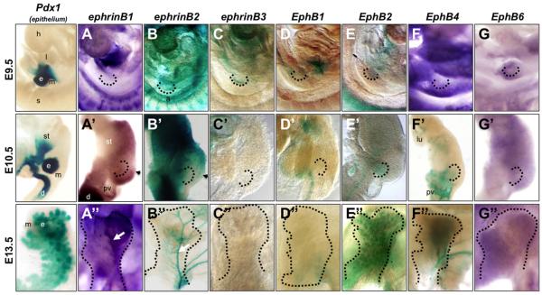
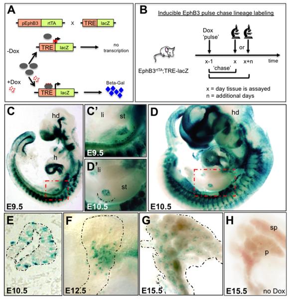
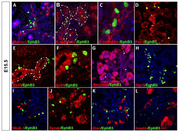
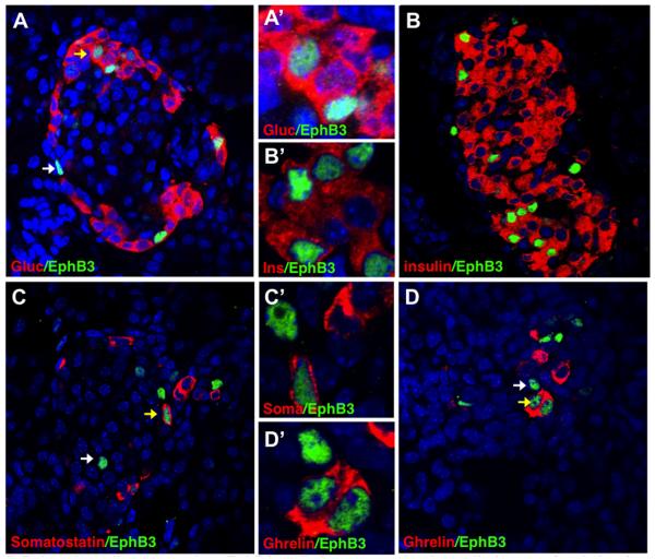
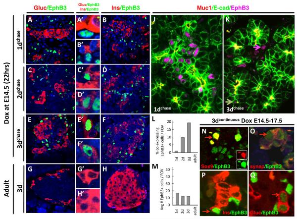
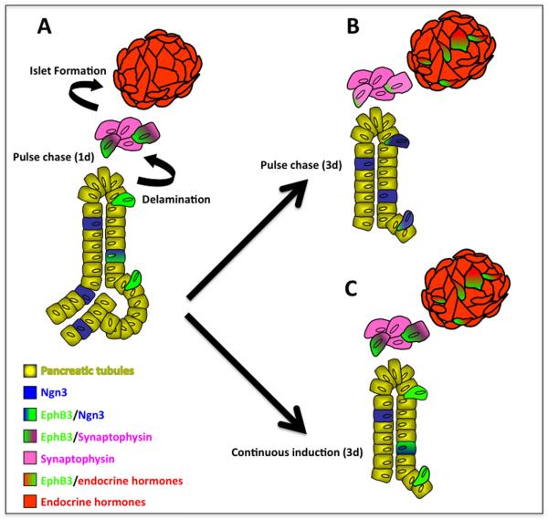
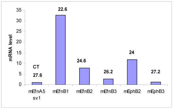
Results: Here, we identify EphB3 as a novel pro-endocrine marker and use its expression to track delaminating islet lineages. First, we provide a detailed developmental expression profile for EphB3 and other EphB family members in the embryonic pancreas. We demonstrate that EphB3 transiently marks endocrine cells as they delaminate from the pancreatic epithelium, prior to their differentiation. Using a Tet-inducible EphB3(rtTA-lacZ) reporter line, we show that short-term pulse-labeled EphB3(+) cells co-express Pdx1, Nkx6.1, Ngn3, and Synaptophysin, but not insulin, glucagon, or other endocrine hormones. Prolonged labeling tracks EphB3(+) cells from their exit from the epithelium to their differentiation.

Conclusions: These studies demonstrate that pro-endocrine cell differentiation during late gestation, from delamination to maturation, takes approximately 2 days. Together, these data introduce EphB3 as a new biomarker to identify beta-cells at a critical step during their step-wise differentiation and define the timeframe of endocrine differentiation.


Figures

Figure 1
Figure 1. Expression of ephrin ligands and Eph receptors during pancreatic development
Whole mount in situ hybridization and whole mount β-galactosidase staining of ephrinB ligands and EphB receptors. First column: β-galactosidase staining of Pdx1-lacZ embryonic midgut and pancreas at stages indicated; epithelium stains blue. In situ hybridization of ephrinB1 (A,A”); EphB4 (F); and EphB6 (G,G”). Whole mount β-galactosidase staining of ephrinB2 (B,B”); ephrinB3 (C,C”); EphB1 (D,D”); EphB2 (E,E”); and EphB4 (F’,F”) at stages indicated. E9.5 embryos in (A-G) are facing forward. E10.5 dissected gut tubes in (A’-G’) have anterior up and dorsal to the right. E13.5 Dorsal pancreatic buds in (A”-G”) have stomach in the background and are shown in lateral view (proximal at bottom, distal at top). The pancreatic endoderm is delineated by dotted black lines. Arrowheads in A’ and B’ show boundary between reciprocal expression domains of ephrinB2 and ephrinB1. White arrows point to ridge (A”) or to main blood vessel (B”). a, aorta; d, duodenum; e, pancreatic epithelium; h, heart; l, liver; lu, lung; m, mesoderm/mesenchyme; pv, portal vein; s, somites; st, stomach.
Figure 2
Figure 2. EphB3rtTA is expressed in the pancreatic anlage
(A) Diagram showing the generation of BAC-Tg-EphB3rtTA-lacZ males. (B) Schematic exemplifying the strategy for EphB3 lineage tracing, either short or long term. (C,D) Whole mount β-galactosidase staining of E9.5 and E10.5 embryos (C’,D’ show close ups of gut tube in C,D, red dotted boxes). (E,F) Whole mount β-galactosidase staining of E12.5 and E15.5 pancreata. Distal is up, proximal is down. (G) Sagittal section of E10.5 pancreatic bud showing expression of EphB3 in scattered cells. (H) E15.5 uninduced pancreas showing no leakage of β-gal expression. Broken lines delineate pancreas. h, heart; hd, head.
Figure 3
Figure 3. Pulse-chase lineage tracing: First transition induction and assay after 1 day (at E9.5 or E10.5)
Immunostaining of anti-β-gal (green) and pancreatic markers (red) show that EphB3 is expressed in the early pancreatic epithelium. (A) Transverse section of E9.5 EphB3rtTA-lacZ pancreatic bud. EphB3+ cells (green) and Pdx-1+ cells (red). (B-F) Sagittal sections of E10.5 pancreata. EphB3 expression (green) and (B) E-cadherin, (C) Sox9, (D) Nkx6.1, (E) glucagon (red) and (F) Ngn3. Arrows indicate overlap. Asterisk indicate lack of overlap in panel C.
Figure 4
Figure 4. Pulse-chase lineage tracing: Second transition induction and assay after 1 day (at E15.5)
Immunostaining of anti-β-gal (green) and pancreatic markers (red) show that EphB3 is expressed in delaminating pro-endocrine cells. Sagittal sections of E15.5 EphB3rtTA-lacZ pancreata. EphB3+ cells (green) and (A) Mucin+, (B,C) E-cadherin+; (D) Pdx1+; (E,F) Sox9+; (G) amylase+; (H) Ngn3+; (I) synaptophysin+ (Synap); (J) Nkx6.1+; (K) glucagon+ (Gluc); or (L) insulin+ (Ins) cells (red). Pancreatic epithelium is outlined by stippled lines; endocrine clusters in B, *.
Figure 5
Figure 5. Pulse-chase lineage tracing: EphB3+ cells become endocrine cells (assay after 8 days)
Dox pulse administered at E14.5 and pancreas assayed at P4. Immunostainings of postnatal EphB3rtTA-lacZ pancreata with anti-β-gal (green) and pancreatic markers (red) show that, following an 8 day chase, EphB3+ cells express (green): (A,A’) glucagon, (B,B’) insulin, (C,C’) somatostain (Sst), or (D,D’) ghrelin (red). Yellow arrows point to overlaps, while white arrows point to EphB3+ cells that do not overlap.
Figure 6
Figure 6. Timeframe of endocrine differentiation of EphB3+ cells: 2 days
Dox pulse is administered at E14.5 for 20 hours, and pancreas is assayed 1 day later (E15.5) (A-B’); 2 days later (E17.5) (C-D’); or 3 days later (E18.5) (E-F’). Alternatively, Dox is administered continuously for 3 days (G-H’). (A-H) Panels show EphB3 co-expression with endocrine hormones: EphB3+ cells (green) and glucagon+ or insulin+ cells (red) are shown in adjacent columns (close ups in center column). Note lack of co-expression of EphB3 and hormones after 1 day pulse-chase, but a few cells co-express after 2 days, and more cells label after 3 days (A,B). Immunostains show pre-ductal epithelium and EphB3+ cells after 1 day of induction (J) and after 2 days (K): Muc1 marks apical side of ductal epithelium (red), E-cadherin marks epithelial cells (green) and EphB3+ (magenta). Graphs show the number of EphB3+ cells co-expressing hormonal markers (insulin and glucagon) (L) and total number of endocrine versus EphB3+ cells per field of view (FOV) (M), at different timepoints post-induction. (N-Q) Stainings of 3 day continuous Dox induction of EphB3rtTA-lacZ: EphB3+ (green) and (N) Sox9+, (O) synapthophysin+; (P) insulin+; or (Q) glucagon+ (red).
Figure 7
Figure 7. Model for EphB3-expressing cell contribution to islets
Following the secondary transition, EphB3 is expressed in a continuum, including three key cell populations during the secondary transition: 1) pro-endocrine cells within the epithelium, 2) ‘delaminating’ pro-endocrine cells, and 3) pro-endocrine cells in early clusters and islets. (A) Short term labeling of EphB3+ cells shows that EphB3 is expressed in delaminating pro-endocrine cells. (B) Pulse-chase experiments determined that EphB3-expressing cells labeled at E14.5 begin to express endocrine hormones by E16.5 and contribute to islets. Newly emerging pro-endocrine cells are labeled due to residual systemic Dox (following a 20 hour pulse of Dox and 8 day chase, expression of EphB3 is restricted to endocrine cells, not shown here). (C) Continuous 3 day Dox induction of EphB3rtTA-lacZ mice labels all endocrine populations, during their step-wise differentiation, from their delamination to hormone expression: both newly emerging pro-endocrine cells and older maturing endocrine cells.
Figure 8
Figure 8. Eph/ephrinB are expressed in adult islets expression
Q-PCR from mRna isolated from mice islets. Primers were validated with brabin mRNA. CT was calculated utilizing cyclophilin expression as baseline. Efn-ephrin.

References

    1. Ahlgren U, Pfaff SL, Jessell TM, Edlund T, Edlund H. Independent requirement for ISL1 in formation of pancreatic mesenchyme and islet cells. Nature. 1997;385:257–60. - PubMed
    1. Apelqvist A, Li H, Sommer L, Beatus P, Anderson DJ, Honjo T, Hrabe de Angelis M, Lendahl U, Edlund H. Notch signalling controls pancreatic cell differentiation. Nature. 1999;400:877–81. - PubMed
    1. Cleaver O, MacDonald RJ. Developmental Molecular Biology of the Pancreas. In: Neoptolemos J, et al., editors. Handbook of Pancreatic Cancer. Springer; New York, NY: 2009.
    1. Cowan CA, Yokoyama N, Saxena A, Chumley MJ, Silvany RE, Baker LA, Srivastava D, Henkemeyer M. Ephrin-B2 reverse signaling is required for axon pathfinding and cardiac valve formation but not early vascular development. Dev Biol. 2004;271:263–71. - PubMed
    1. D’Amour KA, Bang AG, Eliazer S, Kelly OG, Agulnick AD, Smart NG, Moorman MA, Kroon E, Carpenter MK, Baetge EE. Production of pancreatic hormone-expressing endocrine cells from human embryonic stem cells. Nat Biotechnol. 2006;24:1392–401. - PubMed

Publication types

MeSH terms

LinkOut - more resources