. 2012 Jul;40(13):6158-73.
doi: 10.1093/nar/gks231. Epub 2012 Mar 19.

Identification and classification of bacterial Type III toxin-antitoxin systems encoded in chromosomal and plasmid genomes

Affiliations

Identification and classification of bacterial Type III toxin-antitoxin systems encoded in chromosomal and plasmid genomes

Tim R Blower et al. Nucleic Acids Res. 2012 Jul.

Abstract

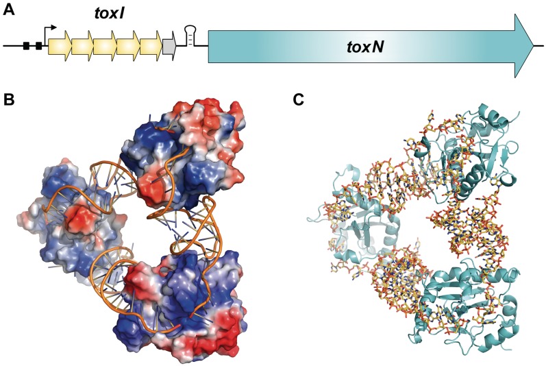
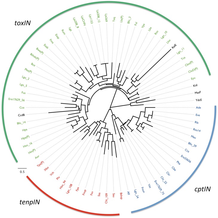
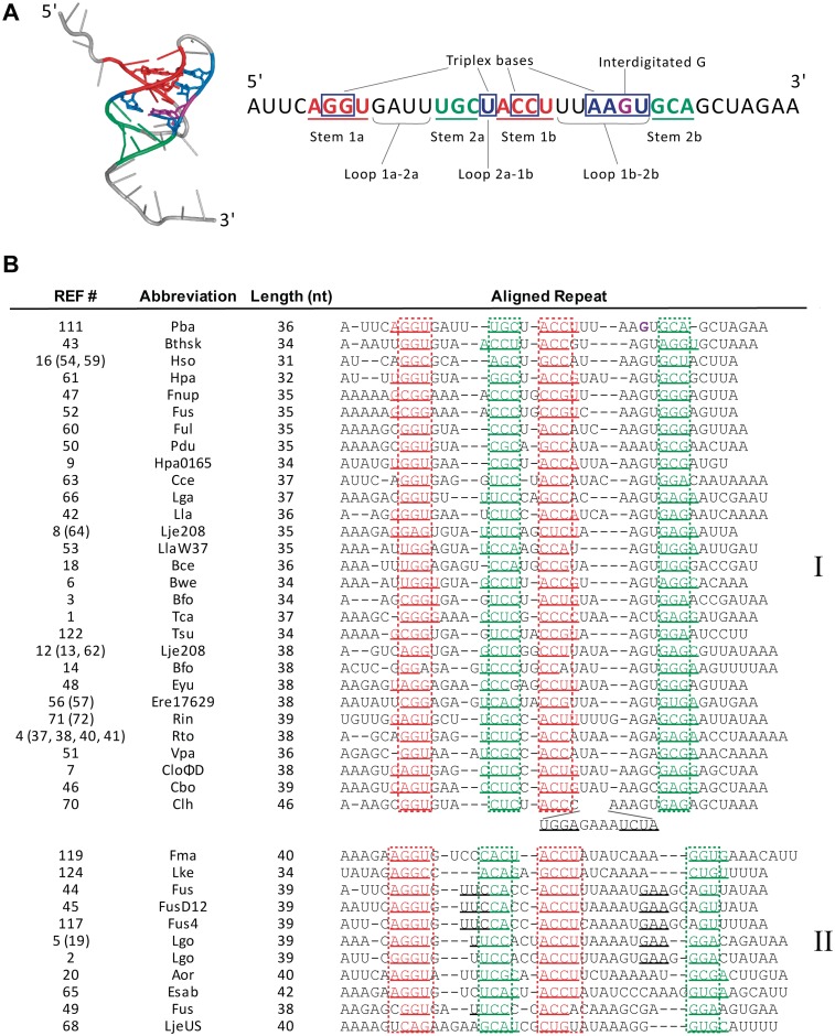
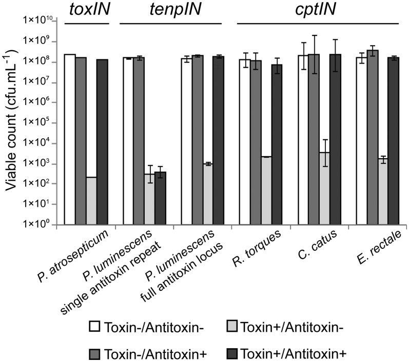
Toxin-antitoxin systems are widespread in bacteria and archaea. They perform diverse functional roles, including the generation of persistence, maintenance of genetic loci and resistance to bacteriophages through abortive infection. Toxin-antitoxin systems have been divided into three types, depending on the nature of the interacting macromolecules. The recently discovered Type III toxin-antitoxin systems encode protein toxins that are inhibited by pseudoknots of antitoxic RNA, encoded by short tandem repeats upstream of the toxin gene. Recent studies have identified the range of Type I and Type II systems within current sequence databases. Here, structure-based homology searches were combined with iterative protein sequence comparisons to obtain a current picture of the prevalence of Type III systems. Three independent Type III families were identified, according to toxin sequence similarity. The three families were found to be far more abundant and widespread than previously known, with examples throughout the Firmicutes, Fusobacteria and Proteobacteria. Functional assays confirmed that representatives from all three families act as toxin-antitoxin loci within Escherichia coli and at least two of the families confer resistance to bacteriophages. This study shows that active Type III toxin-antitoxin systems are far more diverse than previously known, and suggests that more remain to be identified.


Figures

Figure 1.
Figure 1.
Overview of Type III toxin–antitoxin loci. (A) Schematic of a Type III toxin–antitoxin locus. The paradigmatic Type III system, ToxIN from P. atrosepticum, is depicted. The toxin gene, ToxN, is preceded by a stem–loop structure formed from a palindromic repeat. This is itself preceded by a set of tandem nucleotide repeats which act as antitoxic RNA pseudoknots. In the case of ToxIN, the antitoxin, ToxI, is encoded by 5.5, 36 nt tandem repeats (yellow arrows for a full repeat, grey arrow for the half repeat). The locus is transcribed from a constitutive promoter, with the −35 and −10 elements shown as black boxes and the transcriptional start site shown by a black arrow. (B) ToxIN trimer (PDB: 2XDD), with ToxI monomers shown as cartoons and ToxN monomers shown with electrostatic surfaces, where red represents electronegative potential and blue is electropositive. Three monomers of ToxN are held at respective corners of a heterohexameric triangular assembly, formed entirely through protein–RNA interactions with the interspersed pseudoknots of 36 nt ToxI RNAs. (C) ToxIN trimer (PDB: 2XDD) with ToxI shown as yellow sticks and ToxN as cyan cartoons. Figure and legend adapted with the author’s permission from Ref. (4).
Figure 2.
Figure 2.
Taxonomy of Type III toxin–antitoxin loci. The taxonomic distribution of the identified members from each Type III toxin–antitoxin family is shown as a pie chart, with the outer ring representing the different Phyla and the inner portion representing the respective subdivisions of each Phylum into Orders. Class has been omitted for clarity. Colours are as indicated in the inset key.
Figure 3.
Figure 3.
Phylogeny of selected toxin sequences. Sixty-nine toxin sequences from loci unambiguously containing all features of a Type III system (presence of putative antitoxic repeats, promoter, terminator) were aligned, together with five Type II toxins, and then analysed with TREEFINDER (29). In the case where a certain species has more than one Type III TA system selected in this manner, the number following the underscore (e.g. Lgo_14) refers to the reference number for that system (Table 1 and Supplementary Table S1). A ‘P’ in parentheses implies that the source TA system is encoded upon a plasmid, rather than within the chromosome. Entries from the toxIN family are coloured green, cptIN are blue and tenpIN are red, while Type II toxins are in black. The scale bar represents the approximate number of changes per amino acid position as the tree expands radially.
Figure 4.
Figure 4.
Alignment of putative pseudoknot elements with toxIN antitoxins. (A). Structure of a single pseudoknot repeat of P. atrosepticum ToxI (left, PDB: 2XDD), and schematic of secondary and tertiary interactions within each ToxI unit (right). (B) Alignment of consensus repeat sequences of the ToxIN family antitoxins. Stem loops 1 and 2 are shown in red and teal, respectively; additional potential base pairing regions are underlined. The intercalated G19 of ToxI is shown in purple. The reference numbers in brackets indicate entries with identical ToxI consensus repeat sequences, as listed in Supplementary Table S1. Entries 10, 11 and 30 could not be aligned because of overall length (∼60 nt). Entries 15 (69), 17 (39), 55, 58, 67, 118, 120 (121), 123 and 125 all contained two nested base pairing regions of >3 nt each, but could not be aligned, because the loop lengths between base pairing regions did not match the pattern of either group I or II. Strain abbreviations can be related back to entries in Table 1 and Supplementary Table S1.
Figure 5.
Figure 5.
Protection of E. coli DH5α from Type III toxins by cognate antitoxins. Protection assays were performed as described in Materials and Methods. Results for the toxIN system of P. atrosepticum have been published previously (9); data from a single toxIN experiment is included for illustrative purposes. Of the four new loci tested, all toxin genes reduced viability of the host E. coli, which could then be restored by the full cognate antitoxin. Data shown are the mean values from triplicate experiments, with standard deviations represented by error bars.

References

    1. Hayes F. Toxins-antitoxins: plasmid maintenance, programmed cell death, and cell cycle arrest. Science. 2003;301:1496–1499. - PubMed
    1. Gerdes K, Christensen SK, Lobner-Olesen A. Prokaryotic toxin-antitoxin stress response loci. Nat. Rev. Microbiol. 2005;3:371–382. - PubMed
    1. Hayes F, Van Melderen L. Toxins-antitoxins: diversity, evolution and function. Crit. Rev. Biochem. Mol. Biol. 2011;46:386–408. - PubMed
    1. Blower TR, Salmond GP, Luisi BF. Balancing at survival’s edge: the structure and adaptive benefits of prokaryotic toxin-antitoxin partners. Curr. Opin. Struct. Biol. 2011;21:109–118. - PubMed
    1. Leplae R, Geeraerts D, Hallez R, Guglielmini J, Dréze P, Van Melderen L. Diversity of bacterial type II toxin-antitoxin systems: a comprehensive search and functional analysis of novel families. Nucleic Acids Res. 2011;39:5513–5525. - PMC - PubMed

Publication types