. 2012 May 23;134(20):8455-67.
doi: 10.1021/ja211308s. Epub 2012 May 8.

Chemoenzymatic reversible immobilization and labeling of proteins without prior purification

Affiliations

Chemoenzymatic reversible immobilization and labeling of proteins without prior purification

Mohammad Rashidian et al. J Am Chem Soc. .

Abstract

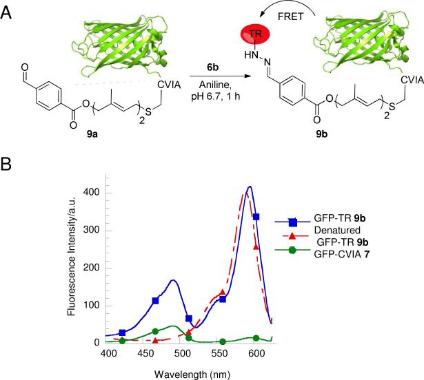
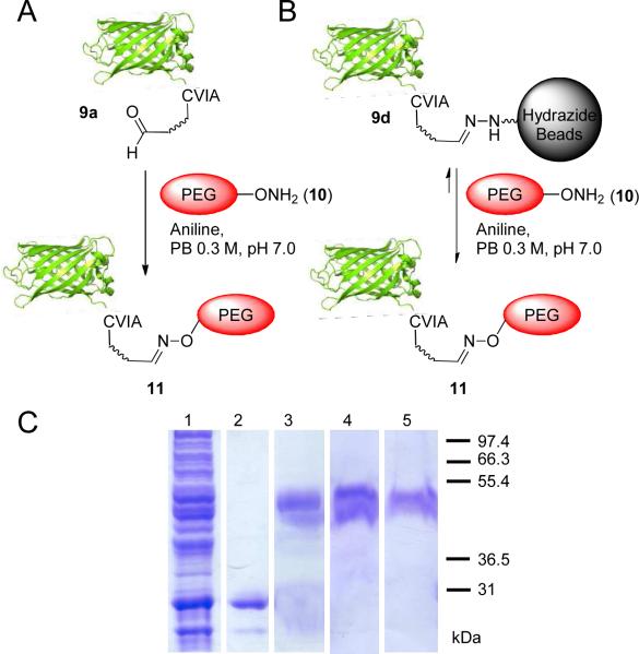
Site-specific chemical modification of proteins is important for many applications in biology and biotechnology. Recently, our laboratory and others have exploited the high specificity of the enzyme protein farnesyltransferase (PFTase) to site-specifically modify proteins through the use of alternative substrates that incorporate bioorthogonal functionality including azides and alkynes. In this study, we evaluate two aldehyde-containing molecules as substrates for PFTase and as reactants in both oxime and hydrazone formation. Using green fluorescent protein (GFP) as a model system, we demonstrate that the purified protein can be enzymatically modified with either analogue to yield aldehyde-functionalized proteins. Oxime or hydrazone formation was then employed to immobilize, fluorescently label, or PEGylate the resulting aldehyde-containing proteins. Immobilization via hydrazone formation was also shown to be reversible via transoximization with a fluorescent alkoxyamine. After characterizing this labeling strategy using pure protein, the specificity of the enzymatic process was used to selectively label GFP present in crude E. coli extract followed by capture of the aldehyde-modified protein using hydrazide-agarose. Subsequent incubation of the immobilized protein using a fluorescently labeled or PEGylated alkoxyamine resulted in the release of pure GFP containing the desired site-specific covalent modifications. This procedure was also employed to produce PEGylated glucose-dependent insulinotropic polypeptide (GIP), a protein with potential therapeutic activity for diabetes. Given the specificity of the PFTase-catalyzed reaction coupled with the ability to introduce a CAAX-box recognition sequence onto almost any protein, this method shows great potential as a general approach for selective immobilization and labeling of recombinant proteins present in crude cellular extract without prior purification. Beyond generating site-specifically modified proteins, this approach for polypeptide modification could be particularly useful for large-scale production of protein conjugates for therapeutic or industrial applications.


Figures

Figure 1
Figure 1
A) Structures of farnesyl diphosphate, farnesyl aldehyde diphosphate (1) and formylbenzoyloxy geranyl diphosphate (2). B) Schematic representation of prenylation of a protein contaning a CAAX-box positioned at its C-terminus (GFP-CVIA, 7) with aldehyde-containing analogue 2 to yield the prenylated product 9a. C) ESI MS analysis of 7 with the deconvoluted mass spectrum shown in the inset. D) ESI MS analysis of 9a with the deconvoluted mass spectrum shown in the inset.
Figure 2
Figure 2
A) Schematic representation of oxime and hydrazone ligations of 8a to yield 8b and 8c. B) Fluorescence (right) and Coomassie blue staining (left) images of a gel loaded with 8a labeled with alexafluor 6c and Texas red 6b via oxime and hydrazone ligations, respectively, showing covalent attachment of fluorophores to the protein. lane 1: GFP-CVIA 7; lane 2: 8c; lane 3: 8b C, D and E) ESI MS spectra of 8a (spectrum C) and hydrazone/oxime ligation products 8b and 8c, showing full conversion for oxime (spectrum D) and ~20% for hydrazone (spectrum E) ligations with the deconvoluted mass spectra shown in the insets.
Figure 3
Figure 3
A) Schematic representation of the fluorescent labeling of 9a via hydrazone ligation. The conjugated protein was expected to show FRET between Texas red and GFP-CVIA. B) Excitation spectra obtained by monitoring at 640 nm. Squares: 9b; Triangles: denatured 9b; Circles: 7; all three samples had equal concentrations of the chromophores.
Figure 4
Figure 4
A) Schematic representation of the fluorescence labeling of 9a via oxime ligation. The conjugated protein was expected to show FRET between TAMRA and GFP. B) Emission spectra obtained by excitation at 488 nm. Circles: 9e; Triangles: GFP-CVIA (7); Squares: 6d; all three samples had equal concentrations of the chromophores. C) Molecular model of GFP–TAMRA 9e conjugate.
Figure 5
Figure 5
A) Schematic representation of immobilization of 9a onto hydrazide functionalized agarose beads to yield 9d. B) Kinetic analysis of immobilization of 9a onto hydrazide functionalized agarose beads. The reaction was carried out at rt, in the presence of 100 mM aniline and excess beads. UV absorbance of GFP in the supernatant was measured at different times showing >95% immobilization in ~45 min. The data was fit to a simple exponential process.
Figure 6
Figure 6
Immobilization onto and subsequent release of 9a from hydrazide-functionalized agarose beads: A) immobilization reaction mixture in the presence of aniline, B) release of 9d from agarose beads via oxime ligation with hydroxylamine in the presence of aniline for ~3 h, and C) control immobilization reaction containing unmodified GFP-CVIA 7. The immobilization reaction was carried out in the presence of protein (54 μM), aniline (100 mM) and PB (100 mM, pH 7). Release of hydrazone-GFP 9d from agarose beads was carried out in the presence of hydroxylamine (200 mM), aniline (100 mM) and PB (200 mM, pH 7). Bright-field images are on the top and fluorescent microscope images are on the bottom. Scale bars in the lower right-hand corners represent 200 μm.
Figure 7
Figure 7
A) Schematic representation of the release of immobilized GFP 9d to yield 9e from agarose beads via oxime ligation with hydroxylamine. B) Kinetic analysis of the release of 9e from agarose beads by oxime ligation. The reaction was carried out at rt, in the presence of 100 mM aniline and 200 mM of hydroxylamine. UV absorbance of GFP in the supernatant was measured over time, which showed approximately 80% release of 9e in 3 h. Analysis of the hydrolytic stability of 9d in the absence of hydroxylamine and aniline showed no detectable release of GFP on the same time scale. The data was fit to a simple exponential decay process.
Figure 8
Figure 8
Chemoenzymatic site-specific tagging of proteins by aldehyde-FPP analogs by PFTase followed by capture of the aldehyde-functionalized protein in the crude cell lysate via hydrazide functionalized beads. Prenylation in the crude extract was confirmed by LC-MS analysis. The immobilized protein was then released into the solution or fluorescently labeled by addition of hydroxylamine or an aminooxy-fluorophore, using aniline as the catalyst. SDS-PAGE analysis: lane 1: crude E. coli lysate containing 9a visualized by Coomassie blue staining; lane 2: 9c released from hydrazide beads after treatment with 6c and visualized by Coomassie blue staining; lane 3: 9c released from hydrazide beads after treatment with 6c and visualized by in gel fluorescence analysis.
Figure 9
Figure 9
A) Generation of site-specifically C-terminal PEGylated GFP from pure 9a. B) MALDI analysis of PEGylated GFP 11. The lower panel is the MALDI spectrum of pure PEG 10, the middle panel is the MALDI spectrum of pure 9a and the top panel is the MALDI spectrum of the oxime PEGylated GFP 11, which confirms complete conversion. The reaction was performed using 9a (10 μM) and 10 (100 μM) for 2 h. Excess of 10 was removed via a zip-tip protocol prior to MALDI analysis.
Figure 10
Figure 10
Use of PFTase-catalyzed protein modification for site-specific PEGylation from purified protein or crude cell lysate. A) Generation of site-specific C-terminal PEGylated protein from pure 9a. B) PEGylation and release of immobilized 9d from hydrazide beads using PEG 10. C) SDS PAGE analysis of PEGylated GFP (11) from purified 9a or from immobilized protein 9d. In case of the crude cell lysate, 7 was chemoenzymatically and site-specifically tagged by aldehyde-containing analog 2 via PFTase catalyzed reaction, followed by capture of the resulting aldehyde-functionalized protein from the lysate using hydrazide functionalized beads. The immobilized protein was then released back into solution and simultaneously site-specifically PEGylated by addition of aminooxy-PEG 10, using aniline as a catalyst. SDS-PAGE analysis: lane 1: crude E. coli lysate containing 9a; lane 2: purified 9a; lane 3: 11 produced by PEGylation of pure 9a with 10; lane 4: 11 prepared from 9d (obtained using purified 9a) and subsequently released with 10; lane 5: 11 prepared from 9d (obtained using 9a present in crude lysate) and subsequently released with 10.
Figure 11
Figure 11
A) Schematic representation of prenylation of glucose-dependent insulinotropic polypeptide (GIP) containing a CAAX-box positioned at its C-terminus (GIP-CVIM, 12a) with aldehyde-containing analogue 2 to yield the prenylated product 12b, which is then site-specifically PEGylated using a short chain aminooxy-PEG (13). B) MALDI MS analysis of prenylation and PEGylation of GIP 12a. MALDI MS spectra (from the top to the bottom) of oxime PEGylated GIP 14, the prenylated aldehyde labeled GIP 12b, and pure 12a, respectively.
Figure 12
Figure 12
Use of PFTase-catalyzed protein modification for site-specific PEGylation of GIP 12a from crude cell lysate. A) Chemoenzymatic site-specific tagging of GIP 12a by aldehyde-FPP analog 2 in the crude cell lysate via PFTase followed by capture of the aldehyde-functionalized polypeptide 12b via hydrazide functionalized beads. The immobilized polypeptide was then released back into the solution and simultaneously site-specifically PEGylated by addition of aminooxy-PEG 13. B) MALDI analysis of the released material confirmed the formation and release of the pure PEGylated GIP (14) into the solution.

References

    1. Zhu H, Bilgin M, Bangham R, Hall D, Casamayor A, Bertone P, Lan N, Jansen R, Bidlingmaier S, Houfek T, Mitchell T, Miller P, Dean RA, Gerstein M, Snyder M. Science. 2001;293:2101–2105. - PubMed
    1. Luk Y-Y, Tingey ML, Dickson KA, Raines RT, Abbott NL. J. Am. Chem. Soc. 2004;126:9024–9032. - PubMed
    1. Weinrich D, Lin P-C, Jonkheijm P, Nguyen UTT, Schröder H, Niemeyer CM, Alexandrov K, Goody R, Waldmann H. Angew. Chem. Int. Ed. 2010;49:1252–1257. - PubMed
    1. Zhong M, Fang J, Wei Y. Bioconjugate Chem. 2010;21:1177–1182. - PubMed
    1. Lin P-C, Ueng S-H, Tseng M-C, Ko J-L, Huang K-T, Yu S-C, Adak AK, Chen Y-J, Lin C-C. Angew. Chem. Int. Ed. 2006;45:4286–4290. - PubMed

Publication types

MeSH terms