Review
. 2012 Mar;246(1):95-106.
doi: 10.1111/j.1600-065X.2012.01108.x.

Ubiquitination in signaling to and activation of IKK

Affiliations
Review

Ubiquitination in signaling to and activation of IKK

Zhijian J Chen. Immunol Rev. 2012 Mar.

Abstract

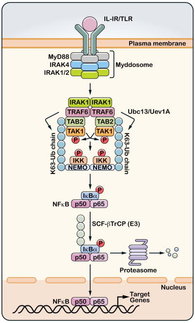
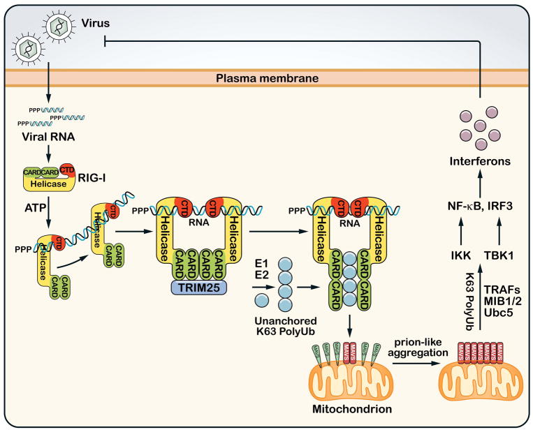
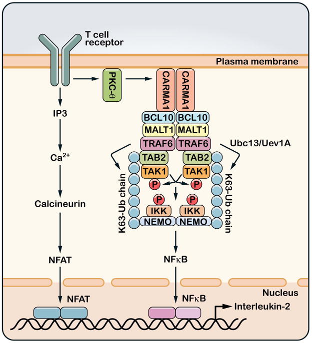
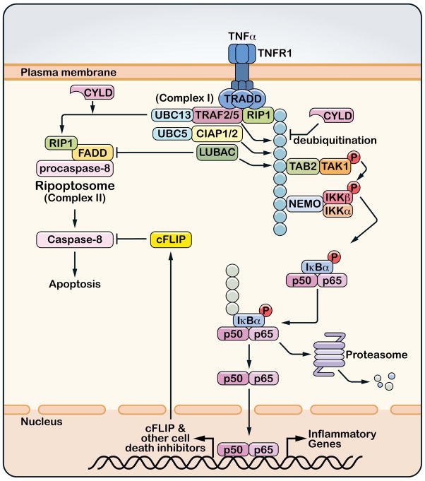
A role for polyubiquitination in the activation of inhibitor of NF-κB (IκB) kinase (IKK) through a proteasome-independent mechanism was first reported in 1996, but the physiological significance of this finding was not clear until 2000 when TRAF6 was found to be a ubiquitin E3 ligase that catalyzes lysine-63 (K63) polyubiquitination. Since then, several proteins known to regulate IKK have been linked to the ubiquitin pathway. These include the deubiquitination enzymes CYLD and A20 that inhibit IKK, and the ubiquitin binding proteins NEMO and TAB2 which are the regulatory subunits of IKK and TAK1 kinase complexes, respectively. Now accumulating evidence strongly supports a central role of K63 polyubiquitination in IKK activation by multiple immune and inflammatory pathways. Interestingly, recent research suggests that some alternative ubiquitin chains such as linear or K11 ubiquitin chains may also play a role in certain pathways such as the TNF pathway. Here I present a historical narrative of the discovery of the role of ubiquitin in IKK activation, review recent advances in understanding the role and mechanism of ubiquitin-mediated IKK activation, and raise some questions to be resolved in future research.


Figures

Figure 1
Figure 1. Ubiquitin-mediated activation of TAK1 and IKK in IL-1R/TLR pathways
Stimulation of interleukin-1 receptor (IL-1R) or one of the Toll-like receptors (TLR) leads to the dimerization of the receptor and subsequent recruitment of the Myddosome complex, which consists of MyD88, IRAK4, IRAK1 (or IRAK2). IRAK1 is phosphorylated by IRAK4 and then associates with and activates the ubiquitin E3 ligase TRAF6. TRAF6 functions together with the ubiquitin E2 complex composed of Ubc13 and Uev1A to catalyze the synthesis of K63-linked polyubiquitin chains which are conjugated to other proteins or unanchored. Unanchored K63 polyubiquitin chains have been shown to bind the TAB2 subunit of the TAK1 kinase complex, and this binding promotes autophosphorylation of TAK1, which results in its activation. The polyubiquitin chains also bind NEMO to recruit the IKK complex, thereby facilitating the phosphorylation of IKKβ by TAK1. IKK is then activated to phosphorylate IκBα, which is polyubiquitinated by the ubiquitin ligase complex SCF-βTrCP and degraded by the 26S proteasome. NF-κB (represented by p50/p65 dimer) translocates to the nucleus to turn on the expression of many target genes.
Figure 2
Figure 2. Ubiquitination in the RIG-I antiviral innate immunity pathway
After infecting a host cell, RNA viruses replicate their RNA, which contains 5′-triphosphate and double-stranded segments that are recognized by the cytosolic sensor protein RIG-I. RIG-I contains an RNA helicase domain and a C-terminal domain (CTD) that bind to viral RNA. The RNA binding and ATP hydrolysis induce the translocation of RIG-I on the RNA and a conformational change that exposes the N-terminal CARD domains, which recruit the ubiquitin E3 TRIM25 to synthesize unanchored K63 polyubiquitin chains. These ubiquitin chains bind to the CARD domains of RIG-I, enabling RIG-I to induce a prion-like aggregation of MAVS on the mitochondrial membrane. The MAVS aggregates then activate IKK and TBK1 through K63 polyubiquitination that involves the E2 Ubc5 and E3s such as TRAFs or MIB1/2. IKK and TBK1 activate NF-κB and IRF3, respectively, which enter the nucleus to induce type-I interferons and other antiviral molecules.
Figure 3
Figure 3. Ubiquitination and T cell activation
Upon engagement of MHC-bound peptides, T cell receptors (TCR) trigger a cascade of tyrosine phosphorylation events that lead to the activation of protein kinase C-θ (PKCθ). PKCθ then phosphorylates the membrane-associated protein CARMA1, which in turn recruits BCL10 and MALT1. MALT1 binds to TRAF6 and perhaps other ubiquitin E3 ligases. The binding of MALT1 to TRAF6 induces TRAF6 oligomerization and activates its E3 ligase activity, which then catalyzes K63 polyubiquitination to activate TAK1 and IKK. T cell receptor signaling also activates the calcineurin – NFAT pathway through increasing the intracellular concentration of calcium. NFAT, NF-κB and other transcription factors cooperate in the nucleus to induce the production of interleukin-2 (IL-2).
Figure 4
Figure 4. Roles of ubiquitination in TNFα-induced NF-κB activation and cell death
The binding of TNFα to its receptor (TNFR1) induces the trimerization of the receptor and recruitment of a protein complex (complex I) that includes the adaptor protein TRADD, the protein kinase RIP1 and ubiquitin E3 ligases TRAF2, TRAF5, cIAP1, cIAP2 and LUBAC. Some or all of these E3s catalyze polyubiquitination of RIP1, which recruits and activates the TAK1 and IKK complexes. Deubiquitination of RIP1 by CYLD not only inhibits TAK1 and IKK activation, but also facilitates the formation of a cytoplasmic complex (complex II or Ripoptosome) consisting of RIP1, FADD and procaspase-8. Within this complex, pro-caspase 8 auto-cleaves to generate mature caspase-8, which then initiates apoptosis. However, caspase-8 is normally inhibited by caspase inhibitors such as c-FLIP, which is induced by NF-κB. Ubiquitin E3 ligases inhibit cell death indirectly by activating NF-κB and/or directly by blocking the formation of Ripoptosome.

References

    1. Hershko A. Ubiquitin: roles in protein modification and breakdown. Cell. 1983;34:11–2. - PubMed
    1. Pickart CM. Ubiquitin enters the new millennium. Mol Cell. 2001;8:499–504. - PubMed
    1. Ciechanover A, Finley D, Varshavsky A. Ubiquitin dependence of selective protein degradation demonstrated in the mammalian cell cycle mutant ts85. Cell. 1984;37:57–66. - PubMed
    1. Finley D, Ciechanover A, Varshavsky A. Thermolability of ubiquitin-activating enzyme from the mammalian cell cycle mutant ts85. Cell. 1984;37:43–55. - PubMed
    1. Scheffner M, Werness BA, Huibregtse JM, Levine AJ, Howley PM. The E6 oncoprotein encoded by human papillomavirus types 16 and 18 promotes the degradation of p53. Cell. 1990;63:1129–36. - PubMed

Publication types

Substances