Review
. 2012 Mar;246(1):154-67.
doi: 10.1111/j.1600-065X.2012.01093.x.

The atypical PKCs in inflammation: NF-κB and beyond

Affiliations
Review

The atypical PKCs in inflammation: NF-κB and beyond

Maria T Diaz-Meco et al. Immunol Rev. 2012 Mar.

Abstract

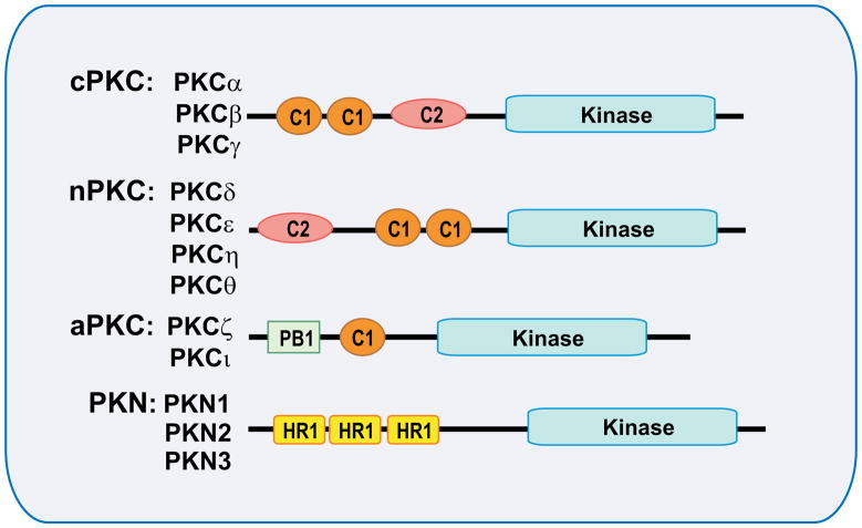
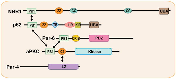
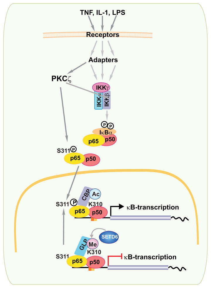
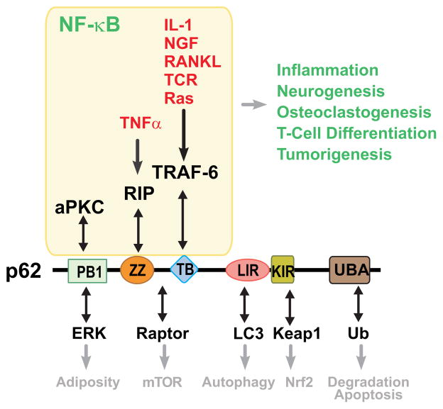
From the very early days of nuclear factor-κB (NF-κB) research, it was recognized that different protein kinase C (PKC) isoforms might be involved in the activation of NF-κB. Pharmacological tools and pseudosubstrate inhibitors suggested that these kinases play a role in this important inflammatory and survival pathway; however, it was the analysis of several genetic mouse knockout models that revealed the complexity and interrelations between the different components of the PB1 network in several cellular functions, including T-cell biology, bone homeostasis, inflammation associated with the metabolic syndrome, and cancer. These studies unveiled, for example, the critical role of PKCζ as a positive regulator of NF-κB through the regulation of RelA but also its inflammatory suppressor activities through the regulation of the interleukin-4 signaling cascade. This observation is of relevance in T cells, where p62, PKCζ, PKCλ/ι, and NBR1 establish a mesh of interactions that culminate in the regulation of T-cell effector responses through the modulation of T-cell polarity. Many questions remain to be answered, not just from the point of view of the implication for NF-κB activation but also with regard to the in vivo interplay between these pathways in pathophysiological processes like obesity and cancer.


Conflict of interest statement

The authors have no conflicts of interest to declare.

Figures

Fig. 1
Fig. 1. Structure of the protein kinase C (PKC)
Schematic representation of the different PKC subfamilies and their domain structural organization. The PKC family is divided into four structurally and functionally distinct subgroups according to their regulatory domains: the classical isoforms (cPKC), novel isoforms (nPKC), atypical isoforms (aPKC) and the PKC-related kinases (PKN). Conserved region 1 (C1) confers binding to diacylglycerol and phospholipids, and C2 senses calcium. PB1 (Phox/Bem domain 1) is specific of aPKC and acts as a dimerization domain. Homology region 1 (HR1) confers small-GTPase binding properties to PKN.
Fig. 2
Fig. 2. The atypical protein kinase C (aPKC) signaling platform
Schematic showing domain organization and network signaling mediated by aPKC and their adapters and regulators. The aPKCs interact with the PB1-containing adapters p62 and Par-6 to regulate specific functions. The scaffold p62 binds NBR1, a highly structurally related molecule with similar domain organization. Par-4 is a regulator and inhibitor of the aPKCs through binding to their C1 domain. C1, conserved region 1; PB1, Phox/Bem domain 1; LZ, leucine zipper; CRIB, Cdc42/Rac interactive binding; PDZ, PSD-95/Dlg/ZO-1; ZZ, ZZ-type zinc finger; TB, TRAF6-binding; LIR, LC3-interacting region; KIR, Keap-interacting region; UBA, ubiquitin-associated; CC, coiled coil.
Fig. 3
Fig. 3. Role of PKCζ in NF-κB activation
The binding of different ligands to their respective receptors in the plasma membrane triggers the recruitment of specific adapters for each receptor that orchestrate the formation of a signalosome complex that includes two catalytic (IKKα and IKKβ) and one regulatory subunit (IKKγ). This complex phosphorylates IκB, which is subsequently ubiquitinated and degraded through the proteasome system, releasing NF-κB (the more classical components of which are p65-p50 heterodimers), which is free now to translocate to the nucleus and interact with elements in the promoter of inflammatory and survival genes harboring κB-elements in their promoters. PKCζ phosphorylates p65 at Serine 311 (S311), an important residue for recruiting the CBP coactivator complex. This event promotes acetylation of Lysine 310 (K310) to activate transcription. Under basal conditions, p65 is methylated (Me) at K310 by SETD6, promoting recruitment of GLP, which leads to the repression of κB-dependent transcription. Upon ligand binding, phosphorylation of S311 by PKCζ blocks methylation to activate the system. Depending on tissue specificity PKCζ could also act as an IKK kinase.
Fig. 4
Fig. 4. p62 is a signaling hub
Schematic showing domain organization and network signaling mediated by p62. The signaling adapter p62 is a critical mediator of important cellular functions owing to its ability to establish interactions with various signaling intermediaries. p62 plays a key role in NF-κB through specific binding to aPKC, RIP and TRAF6 to control inflammation, neurogenesis, osteoclastogenesis, T-cell differentiation and tumorigenesis. PB1, PB1 dimerization domain; ZZ, ZZ-type zinc finger; TB, TRAF6-binding; LIR, LC3-interacting region; KIR, Keap-interacting region; UBA, ubiquitin-associated.

References

    1. Hanks SK, Hunter T. Protein kinases 6. The eukaryotic protein kinase superfamily: kinase (catalytic) domain structure and classification. FASEB J. 1995;9:576–596. - PubMed
    1. Mellor H, Parker PJ. The extended protein kinase C superfamily. Biochem J. 1998;332:281–292. - PMC - PubMed
    1. Nishizuka Y. The role of protein kinase C in cell surface signal transduction and tumour promotion. Nature. 1984;308:693–698. - PubMed
    1. Nakamura S, Yamamura H. Yasutomi Nishizuka: father of protein kinase C. J Biochem. 2010;148:125–130. - PubMed
    1. Kikkawa U, Takai Y, Tanaka Y, Miyake R, Nishizuka Y. Protein kinase C as a possible receptor protein of tumor-promoting phorbol esters. J Biol Chem. 1983;258:11442–11445. - PubMed

Publication types