. 2012 Sep;18(9):697-709.
doi: 10.1089/ten.TEC.2011.0744. Epub 2012 May 21.

Longitudinal in vivo imaging to assess blood flow and oxygenation in implantable engineered tissues

Affiliations

Longitudinal in vivo imaging to assess blood flow and oxygenation in implantable engineered tissues

Sean M White et al. Tissue Eng Part C Methods. 2012 Sep.

Abstract

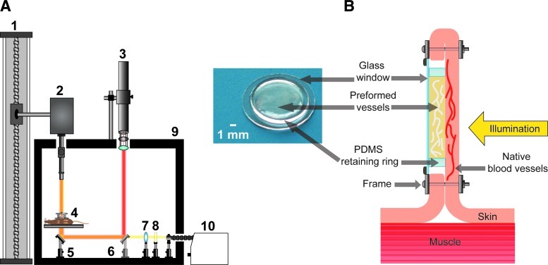
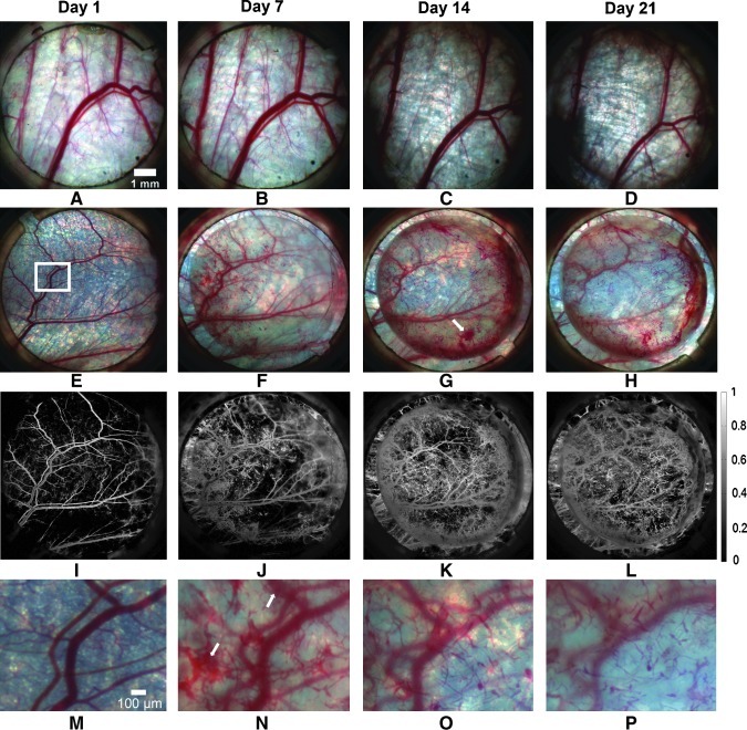
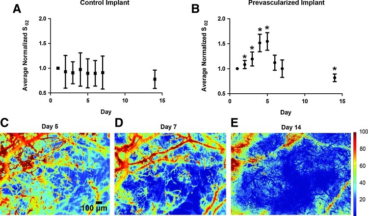
The functionality of vascular networks within implanted prevascularized tissues is difficult to assess using traditional analysis techniques, such as histology. This is largely due to the inability to visualize hemodynamics in vivo longitudinally. Therefore, we have developed dynamic imaging methods to measure blood flow and hemoglobin oxygen saturation in implanted prevascularized tissues noninvasively and longitudinally. Using laser speckle imaging, multispectral imaging, and intravital microscopy, we demonstrate that fibrin-based tissue implants anastomose with the host (severe combined immunodeficient mice) in as short as 20 h. Anastomosis results in initial perfusion with highly oxygenated blood, and an increase in average hemoglobin oxygenation of 53%. However, shear rates in the preformed vessels were low (20.8±12.8 s(-1)), and flow did not persist in the vast majority of preformed vessels due to thrombus formation. These findings suggest that designing an appropriate vascular network structure in prevascularized tissues to maintain shear rates above the threshold for thrombosis may be necessary to maintain flow following implantation. We conclude that wide-field and microscopic functional imaging can dynamically assess blood flow and oxygenation in vivo in prevascularized tissues, and can be used to rapidly evaluate and improve prevascularization strategies.


Figures

FIG. 1.
FIG. 1.
(A) Multimodal imaging platform used to perform laser speckle imaging (LSI) and multispectral imaging (MSI). (1) Motorized slide for large height adjustment of multispectral camera. (2) Multispectral camera with variable magnification lens system and liquid crystal tunable filter that can be removed from the beam path. (3) Helium-neon laser with attached polarizer for intensity attenuation and beam expansion optics used for transillumination during LSI. (4) Mouse bearing dorsal window chamber resting upon circulating water heating block. (5) Mirror at 45° used to redirect light through dorsal window chamber for transillumination. (6) Removable mirror at 45° to redirect laser light for transillumination during LSI. (7) Collimation optics for quartz halogen lamp. (8) Infrared filter. (9) Light-tight enclosure. (10) 150 W quartz halogen lamp. (B) Magnified view of (4) showing mouse dorsal window chamber in cross-section, orientation of implanted prevascularized tissue, and direction of illumination. Inset photograph is a prevascularized tissue, shown with the polydimethylsiloxane (PDMS) ring and 12-mm cover slip present during in vitro culture and implantation into the dorsal window chamber. Color images available online at www.liebertonline.com/tec
FIG. 2.
FIG. 2.
MSI data allow visualization of implanted vessels perfused with blood from host vasculature following anastomosis. Perfused implanted vessels increased in number and are remodeled over the 21-day observation period. Color images of an acellular control implant (A–D) and a prevascularized implant (E–H) over a period of 21 days. (I–L) Hemoglobin absorption maps corresponding to (E–H). (M–P) Magnified view of the region of interest highlighted in (E) over the same time period. White arrows indicate extravasated blood. Color images available online at www.liebertonline.com/tec
FIG. 3.
FIG. 3.
Average hemoglobin oxygenation of prevascularized implants temporarily increases following anastomosis with the host. Plot of average normalized hemoglobin oxygenation values in entire window chamber for control (A) (n=5) and prevascularized implants (B) (n=12) over 14 days. Error bars represent standard deviation among mice at each time point. (C–E) Hemoglobin saturation maps of prevascularized tissue on day 5 (C), day 7 (D), and day 14 (E), demonstrating progressive deoxygenation of preformed vessels. Units are percent hemoglobin oxygen saturation. Asterisk indicates significant (p<0.05 via Mann–Whitney test) change from day 1 value. Color images available online at www.liebertonline.com/tec
FIG. 4.
FIG. 4.
LSI data show that flow does not persist in the majority of blood-filled preformed vessels. (A1–D1) Color images and corresponding speckle flow index (SFI) maps (A2–D2) acquired using LSI of prevascularized implant on days 1, 7, 14, and 21. (E1) Color image of perfused preformed vessels and underlying native vessels on day 7. Blood-filled preformed vessels can be identified by their small diameter and characteristic tortuous morphology (black arrow). (E2) SFI map with T=1000 ms corresponding to (E1) showing that flow is present in native vessels but absent in previously perfused implanted vessels. Unit is s−1. Color images available online at www.liebertonline.com/tec
FIG. 5.
FIG. 5.
Lack of flow in implanted vessels following initial perfusion results in a significant decrease in the ratio of functional vessels to nonfunctional vessels (functional vascular density/vascular density [FVD/VD]). Plots of FVD and FVD/VD for control (A) (n=5) and prevascularized implants (B) (n=12). Ingrowth of only functional vessels in control implants results in a significant increase in VD, but insignificant change in FVD/VD over time. In prevascularized tissues, perfusion followed by thrombosis in implanted vessels results in significant increase in VD without proportional increase in FVD. As a result, FVD/VD decreases significantly over time. Error bars represent standard deviation among mice at each time point. Asterisk indicates significant (p<0.05 via Mann–Whitney test) change from day 1 value. Color images available online at www.liebertonline.com/tec
FIG. 6.
FIG. 6.
Host blood initially perfuses implanted vessels; however, the formation of thrombi prevents continued flow within these vessels. (A) Color image of border between prevascularized implant (below black-dotted line) and native tissue (above black-dotted line). White arrows indicate regions of anastomosis. (B) Fluorescence image of region in (A) indicating that no fluorescent dextran can flow into the prevascularized tissue. (A) and (B) were acquired 5 days following initial implanted vessel perfusion. (C–G) Fluorescent images acquired 30 (C–F) and 40 (G) min post-fluorescein isothiocyanate–dextran injection. There is a 2 s delay between each image (C–F). (C) Shows flowing mouse erythrocytes (white arrows). (D) Shows two preformed vessels where wall shear rate was computed (red arrow=3.2 s−1, yellow arrow=16 s−1). (G) Shows the formation of thrombus (white arrow) that induced flow cessation. (C–G) were acquired <1 h following initial implanted vessel perfusion. Color images available online at www.liebertonline.com/tec

References

    1. Carmeliet P. Jain R.K. Angiogenesis in cancer and other diseases. Nature. 2000;407:249. - PubMed
    1. Muschler G.F. Nakamoto C. Griffith L.G. Engineering principles of clinical cell-based tissue engineering. J Bone Joint Surg Am. 2004;86:1541. - PubMed
    1. Helmlinger G. Yuan F. Dellian M. Jain R.K. Interstitial pH and pO2 gradients in solid tumors in vivo: high-resolution measurements reveal a lack of correlation. Nat Med. 1997;3:177. - PubMed
    1. Liu P. Deng Z. Han S. Liu T. Wen N. Lu W. Geng X. Huang S. Jin Y. Tissue-engineered skin containing mesenchymal stem cells improves burn wounds. Artif Organs. 2008;32:925. - PubMed
    1. Nie X. Cai J.K. Yang H.M. Xiao H.A. Wang J.H. Wen N. Zhang Y.J. Jin Y. Successful application of tissue-engineered skin to refractory ulcers. Clin Exp Dermatol. 2007;32:699. - PubMed

Publication types