. 2012 Oct;16(10):2429-39.
doi: 10.1111/j.1582-4934.2012.01560.x.

Elastin overexpression by cell-based gene therapy preserves matrix and prevents cardiac dilation

Affiliations

Elastin overexpression by cell-based gene therapy preserves matrix and prevents cardiac dilation

Shu-Hong Li et al. J Cell Mol Med. 2012 Oct.

Abstract

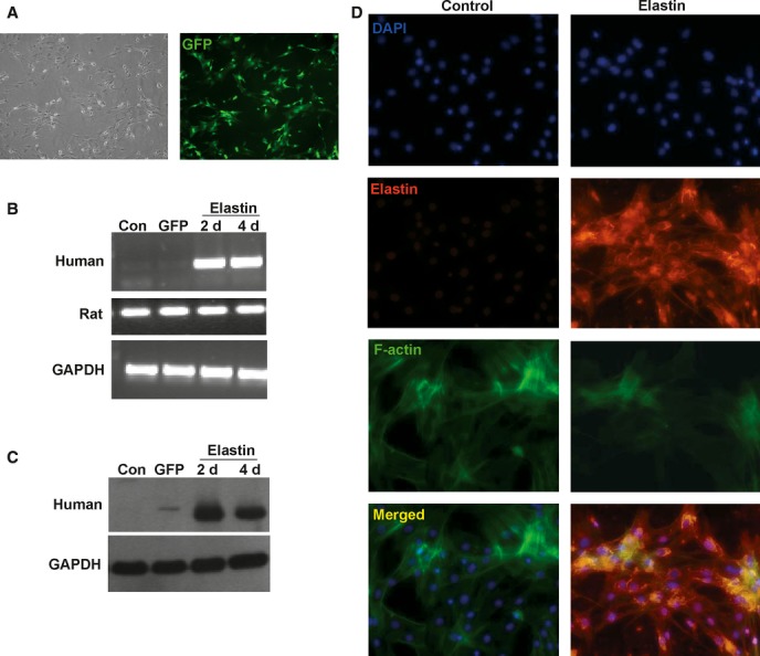
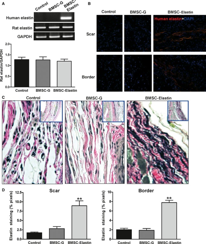
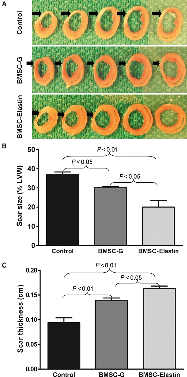
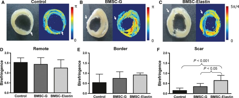
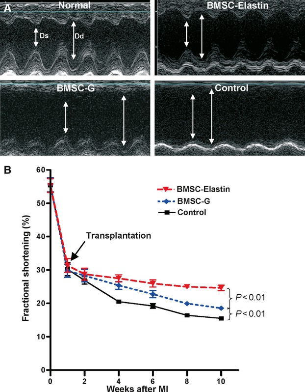
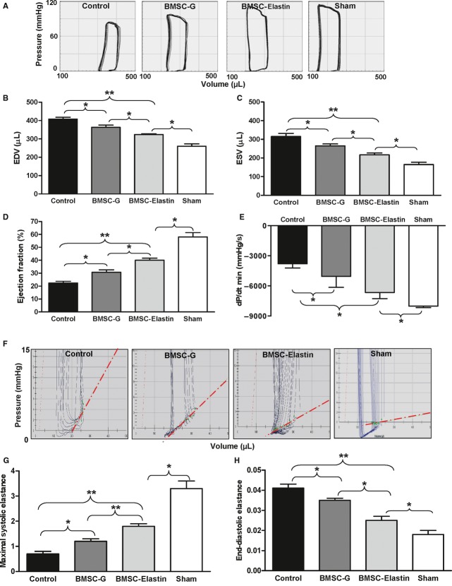
After a myocardial infarction, thinning and expansion of the fibrotic scar contribute to progressive heart failure. The loss of elastin is a major contributor to adverse extracellular matrix remodelling of the infarcted heart, and restoration of the elastic properties of the infarct region can prevent ventricular dysfunction. We implanted cells genetically modified to overexpress elastin to re-establish the elastic properties of the infarcted myocardium and prevent cardiac failure. A full-length human elastin cDNA was cloned, subcloned into an adenoviral vector and then transduced into rat bone marrow stromal cells (BMSCs). In vitro studies showed that BMSCs expressed the elastin protein, which was deposited into the extracellular matrix. Transduced BMSCs were injected into the infarcted myocardium of adult rats. Control groups received either BMSCs transduced with the green fluorescent protein gene or medium alone. Elastin deposition in the infarcted myocardium was associated with preservation of myocardial tissue structural integrity (by birefringence of polarized light; P < 0.05 versus controls). As a result, infarct scar thickness and diastolic compliance were maintained and infarct expansion was prevented (P < 0.05 versus controls). Over a 9-week period, rats implanted with BMSCs demonstrated better cardiac function than medium controls; however, rats receiving BMSCs overexpressing elastin showed the greatest functional improvement (P < 0.01). Overexpression of elastin in the infarcted heart preserved the elastic structure of the extracellular matrix, which, in turn, preserved diastolic function, prevented ventricular dilation and preserved cardiac function. This cell-based gene therapy provides a new approach to cardiac regeneration.


Figures

Fig 1
Fig 1
Recombinant elastin expression in rat BMSCs. (A) Cultured rat BMSCs were effectively transduced with the adenovector, as indicated by GFP fluorescence in Ad-CMV-GFP transduced cells at day 4 (40× magnification). (B) Human elastin mRNA in the BMSCs was evaluated by RT-PCR 2 and 4 days after transduction, with primer sets specific for human and rat elastin. (C) Western blotting with an antibody specific for human tropoelastin confirmed the expression of elastin protein in the Ad-CMV-elastin transduced rat BMSCs. (D) Immunostaining with DAPI, anti-human elastin antibody and FITC-conjugated phalloidin (for F-actin) revealed no detectable human elastin in the culture of BMSCs transduced with empty vector (left panels). However, BMSCs transduced with Ad-CMV-elastin (right panels) showed a network of human elastin molecules (200× magnification). As the strong elastin staining in the Ad-CMV-elastin group overshadowed the F-actin staining, we enhanced the contrast/brightness of the F-actin staining to show that most of the elastin-staining cells were F-actin positive. (Con = empty vector; GFP = Ad-CMV-GFP; representative of three experiments).
Fig 2
Fig 2
Expression of elastin in the infarcted myocardium. (A) Seven days after ligation, BMSCs transduced with Ad-CMV-elastin (BMSC-elastin) or Ad-CMV-GFP (BMSC-G) were injected into the central region and border zone of the infarct. Medium alone served as control. Human and rat elastin mRNA in the infarct and border regions was evaluated by RT-PCR 7 days after injection, with primer sets specific for human and rat elastin. GAPDH served as a loading control. The BMSC-elastin group expressed human elastin mRNA, but no human elastin mRNA was found in the medium control or BMSC-G groups. Induction of the human elastin gene in transduced rat BMSCs did not alter the expression of endogenous elastin as no significant changes in rat elastin gene expression were observed among the medium control (n = 3), BMSC-G (n = 3) or BMSC-elastin (n = 5) groups. (B) Seven days after ligation, BMSCs transduced with Ad-CMV-elastin (BMSC-elastin) were injected into the infarct. Medium alone served as control. Human elastin in the infarct and border regions was evaluated by immunofluorescent staining 7 days after injection. Representative micrographs (200× magnification) show that the BMSC-elastin group expressed human elastin protein, but no human elastin was found in the medium control and BMSC-G groups. Nuclei were stained with DAPI. (C) Verhoeff Van Gieson staining of mid-papillary transverse myocardial sections (400× and 40× magnification) of infarcted myocardium shows elastin fibres in the medium control, BMSC-G and BMSC-elastin groups 9 weeks after cell transplantation. (D) The pixel area positive for elastin was significantly greater in the scar and border zone of the BMSC-elastin group compared with medium control and BMSC-G groups (n = 5 per group for the scar, n = 6 per group for the border zone) (**P < 0.01 versus control).
Fig 3
Fig 3
Infarct scar characterization. (A) Sections of infarcted rat hearts show fibrotic scar tissue in the LV free wall (arrows) 9 weeks after cell transplantation. (B, C) Scar size (n = 5 per group) and thickness (n = 4 per group) were evaluated by computed planimetry. The BMSC-elastin group had the smallest and thickest scars compared with the other two groups.
Fig 4
Fig 4
Tissue organization of the infarcted myocardium. (A–C) Sections of infarcted rat hearts (left panels) were used for birefringence imaging analysis (right panels) 9 weeks after cell transplantation. White arrows indicate scar area. Myocardial birefringence (retardance) was determined by polarized light transmission through tissue sections and provides an index of tissue structural organization. (D–F) Myocardial birefringence (measured in radians) of the infarcted myocardium showed no significant group differences in the remote region (D, n = 3 per group) or border zone (E, n = 3 per group); however, within the infarct scar (F, n = 4 per group), birefringence in the BMSC-elastin group was significantly higher than that in the medium control and BMSC-G groups, indicating maintenance of tissue organization with elastin overexpression.
Fig 5
Fig 5
Echocardiographic analysis of cardiac function. (A) Echocardiographic M-mode traces of rat hearts before MI (normal) and 10 weeks after MI. Ds = end-systolic dimension, Dd = end-diastolic dimension. (B) Prior to and 1 week after MI, there was no difference in fractional shortening among groups (n = 8 per group). However, 10 weeks post-MI, the BMSC-G group showed significant preservation of fractional shortening compared with the control group, whereas the BMSC-elastin group showed significantly greater fractional shortening compared with either the medium control or BMSC-G group.
Fig 6
Fig 6
Pressure–volume analysis of cardiac function. (A) Representative P–V loops of rat hearts 9 weeks after cell transplantation. A sham non-ligated control depicts normal cardiac function. The BMSC-G and BMSC-elastin groups had smaller ventricular volumes than the medium control group. (B, C) The BMSC-elastin group had significantly smaller end-diastolic volume (EDV) and end-systolic volume (ESV) than the BMSC-G group (n = 4 per group). (D, E) The load-dependent index of systolic function (ejection fraction) was significantly greater in the BMSC-elastin group than the BMSC-G group, which was better than the medium control group. Diastolic function (dP/dtmin) was significantly better in the BMSC-elastin group versus the other two groups (n = 5 per group). (F) Representative P–V loops during vena cava occlusion demonstrate diastolic function and the position and slope of the end-diastolic pressure–volume relationship (EDPVR, red dotted line). (G, H) The load-independent indices of both systolic and diastolic function (maximal systolic elastance and end-diastolic elastance) were significantly improved in the BMSC-elastin group compared with the BMSC-G group, which was better than the medium control group (n = 4 per group). Elastin overexpression resulted in a more compliant left ventricle. (*P < 0.05, **P < 0.01).

References

    1. Pfeffer JM, Pfeffer MA, Fletcher PJ, et al. Progressive ventricular remodeling in rat with myocardial infarction. Am J Physiol. 1991;260:H1406–14. - PubMed
    1. Raya TE, Gay RG, Lancaster L, et al. Serial changes in left ventricular relaxation and chamber stiffness after large myocardial infarction in rats. Circulation. 1988;77:1424–31. - PubMed
    1. Kielty CM, Sherratt MJ, Shuttleworth CA. Elastic fibres. J Cell Sci. 2002;115:2817–28. - PubMed
    1. Thurmond F, Trotter J. Morphology and biomechanics of the microfibrillar network of sea cucumber dermis. J Exp Biol. 1996;199:1817–28. - PubMed
    1. Reber-Muller S, Spissinger T, Schuchert P, et al. An extracellular matrix protein of jellyfish homologous to mammalian fibrillins forms different fibrils depending on the life stage of the animal. Dev Biol. 1995;169:662–72. - PubMed

Publication types

MeSH terms