Review
. 2011 Jul;1(3):199-216.
doi: 10.1016/j.dcn.2011.04.001. Epub 2011 Apr 27.

The physiology of developmental changes in BOLD functional imaging signals

Affiliations
Review

The physiology of developmental changes in BOLD functional imaging signals

Julia J Harris et al. Dev Cogn Neurosci. 2011 Jul.

Abstract

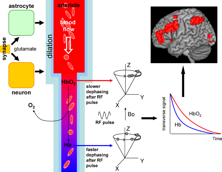
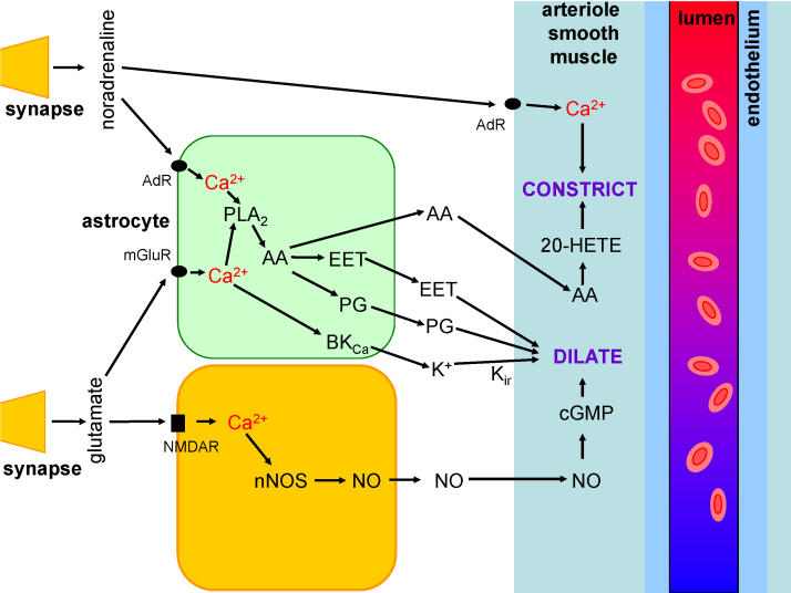
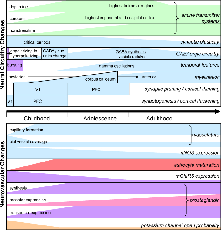
BOLD fMRI (blood oxygenation level dependent functional magnetic resonance imaging) is increasingly used to detect developmental changes of human brain function that are hypothesized to underlie the maturation of cognitive processes. BOLD signals depend on neuronal activity increasing cerebral blood flow, and are reduced by neural oxygen consumption. Thus, developmental changes of BOLD signals may not reflect altered information processing if there are concomitant changes in neurovascular coupling (the mechanism by which neuronal activity increases blood flow) or neural energy use (and hence oxygen consumption). We review how BOLD signals are generated, and explain the signalling pathways which convert neuronal activity into increased blood flow. We then summarize in broad terms the developmental changes that the brain's neural circuitry undergoes during growth from childhood through adolescence to adulthood, and present the changes in neurovascular coupling mechanisms and energy use which occur over the same period. This information provides a framework for assessing whether the BOLD changes observed during human development reflect altered cognitive processing or changes in neurovascular coupling and energy use.


Figures

Fig. 1
Fig. 1
Schematic diagram showing the different stages of how BOLD signals are generated, from neurobiology through physics to data analysis. On the left, neuronal activity releases transmitters (glutamate) which act via neuronal and astrocytic signalling systems to trigger an increase of local blood flow. Neuronal activity also leads to O2 consumption and generation of paramagnetic deoxygenated haemoglobin (Hb) from diamagnetic oxygenated haemoglobin (HbO2). The blood flow increase brings in fresh oxygenated blood which (in adults) lowers the local concentration of Hb. This decreases the non-homogenizing effect that Hb has on the local magnetic field which protons in H2O experience. As a result, after a radiofrequency (RF) pulse is applied transverse to the magnetic field used to align the proton spins (Bo), the synchronised spin precession in the transverse plane dephases more slowly (graph on right). The difference in decay time between the red (HbO2) and blue (Hb) curves in the graph generates the increased MRI signal from protons in areas where neurons are active, which is represented as the red spots superimposed on a structural image of the brain at the top right.
Fig. 2
Fig. 2
Signalling pathways mediating neurovascular coupling. When neurons are active they release glutamate which generates action potentials in interneurons containing NOS and activates metabotropic glutamate receptors (mGluR) in astrocytes. As a result [Ca2+]i rises in both cell types, releasing NO and derivatives of arachidonic acid to dilate local arterioles and increase blood flow. In addition arachidonic acid diffuses to arterioles and is converted into 20-HETE which constricts the vessels. K+ release from astrocytes via large conductance Ca2+-activated K+ channels (BKCa channels) may also dilate vessels by promoting K+ efflux through smooth muscle inward rectifier K+ channels (Kir) and thus generating hyperpolarization. Blood flow changes evoked by release of adenosine, lactate and interneuron peptide transmitters are not shown on this diagram. Vasodilation occurs in the context of constriction of arterioles produced by the amine transmitters noradrenaline, dopamine and 5-HT. Noradrenaline evokes constriction by acting on α-adrenergic receptors on arteriole smooth muscle and on astrocytes. Developmental changes in any of the components will alter the BOLD response.
Fig. 3
Fig. 3
Summary of the developmental time courses of changes in neural information processing mechanisms (top) and of components of the signalling pathways regulating blood flow and thus controlling the BOLD response (bottom). Changes shown in blue are from human data; green is from macaque; lilac from rat or mouse; red from cat; pink from pig; light orange from rabbit.

Comment in

References

    1. Akerman C.J., Cline H.T. Refining the roles of GABAergic signaling during neural circuit formation. Trends Neurosci. 2007;30:382–389. - PubMed
    1. Akgören N., Fabricius M., Lauritzen M. Importance of nitric oxide for local increases of blood flow in rat cerebellar cortex during electrical stimulation. Proc. Natl. Acad. Sci. USA. 1994;91:5903–5907. - PMC - PubMed
    1. Akgören N., Dalgaard P., Lauritzen M. Cerebral blood flow increases evoked by electrical stimulation of rat cerebellar cortex: relation to excitatory synaptic activity and nitric oxide synthesis. Brain Res. 1996;710:204–214. - PubMed
    1. Alkayed N.J., Narayanan J., Gebremedhin D., Medhora M., Roman R.J., Harder D.R. Molecular characterization of an arachidonic acid epoxygenase in rat brain astrocytes. Stroke. 1996;27:971–979. - PubMed
    1. Alle H., Roth A., Geiger J.R. Energy-efficient action potentials in hippocampal mossy fibers. Science. 2009;325:1405–1408. - PubMed

Publication types

MeSH terms