. 2012 Jul 2;169(1):12-27.
doi: 10.1016/j.autneu.2012.02.004. Epub 2012 Mar 20.

Macrophages associated with the intrinsic and extrinsic autonomic innervation of the rat gastrointestinal tract

Affiliations

Macrophages associated with the intrinsic and extrinsic autonomic innervation of the rat gastrointestinal tract

Robert J Phillips et al. Auton Neurosci. .

Abstract

Interactions between macrophages and the autonomic innervation of gastrointestinal (GI) tract smooth muscle have received little experimental attention. To better understand this relationship, immunohistochemistry was performed on GI whole mounts from rats at three ages. The phenotypes, morphologies, and distributions of gut macrophages are consistent with the cells performing extensive housekeeping functions in the smooth muscle layers. Specifically, a dense population of macrophages was located throughout the muscle wall where they were distributed among the muscle fibers and along the vasculature. Macrophages were also associated with ganglia and connectives of the myenteric plexus and with the sympathetic innervation. Additionally, these cells were in tight registration with the dendrites and axons of the myenteric neurons as well as the varicosities along the length of the sympathetic axons, suggestive of a contribution by the macrophages to the homeostasis of both synapses and contacts between the various elements of the enteric circuitry. Similarly, macrophages were involved in the presumed elimination of neuropathies as indicated by their association with dystrophic neurons and neurites which are located throughout the myenteric plexus and smooth muscle wall of aged rats. Importantly, the patterns of macrophage-neuron interactions in the gut paralleled the much more extensively characterized interactions of macrophages (i.e., microglia) and neurons in the CNS. The present observations in the PNS as well as extrapolations from homologous microglia in the CNS suggest that GI macrophages play significant roles in maintaining the nervous system of the gut in the face of wear and tear, disease, and aging.


Figures

Figure 1
Figure 1
Macrophages immunoreactive for CD163 and major histocompatibility complex class II (MHCII) are present in the smooth muscle wall of the gastrointestinal (GI) tract. Macrophages labeled with the antibody to CD163 (A; blue/grey cells) are uniformly distributed throughout the smooth muscle. In contrast, cells labeled with an antibody to MHCII (B; blue/grey cells) are distributed in a highly variable or patchy pattern. The disparate densities of the two phenotypes are illustrated in the two low power montages, each consisting of four images, double labeled for the panneuronal marker human neuronal protein HuC/HuD (HuC/D; brown neurons) and either CD163 (A) or MHCII (B). The two montages are from whole mounts of the small intestine of adult rats and illustrate macrophage distribution patterns in the first third of the rat’s lifespan. Scale bars = 100 µm in A and B.
Figure 2
Figure 2
Monocytes and amoeboid-like cells immunoreactive for CD163 and MHCII are present in the smooth muscle of the GI tract. (A) Monocytes immunoreactive for CD163 have an unstained central nucleus surrounded by heavily stained, granular cytoplasm. (B) Four CD163+ cells with amoeboid-like morphology. (C) A dense cluster of monocyte/amoeboid-like cells and a single bipolar CD163+ macrophage, located above the cells, with two opposing primary processes originating from a centrally located soma. (D) Two CD163+ cells with multiple rudimentary branching processes projecting from their respective somata. In contrast to CD163 labeled monocytes (as in A), MHCII+ monocytes (E) are small and spherical in shape with poorly defined nuclei and cytoplasm. (E) Five monocytes stained for MHCII (blue/grey) surround a single HuC/D+ neuron (brown) in the myenteric plexus. (F) Two MHCII+ monocytes (blue/grey), with poorly developed branching filamentous processes, in close proximity to HuC/D+ myenteric neurons (brown). Focus stacking was used to create an extended depth of field in panel F. Scale bar = 10 µm in F (applies to A–F).
Figure 3
Figure 3
The morphology of macrophages is dynamic and diverse. (A) A single macrophage immunoreactive for CD163 with relatively smooth branching primary processes and blunt or bulbous endings at the tips of its processes. (B–D) Three bipolar macrophages, one immunoreactive for CD163 (B), and two for MHCII (C,D) with numerous thread-like filopodia and nodular outgrowths protruding from the cell’s surface. (E) A stellate macrophage with a morphology similar to the cells in panels B–D. Numerous nodules or spines (F; arrowheads) and thread-like (G) filopodia protrude from the branching processes of the macrophage. Spines and filopodia were often observed on or near the cell body (B–G), but also were found on the primary processes extending away from the macrophage soma (H,I). Two macrophages are shown in panels H and I with multiple long, thick processes that project away from the cell’s body into the surrounding smooth muscle. Images are from whole mounts of the stomach (B) and small intestine (A, C–I) of adult (C–G,I) and aged (A,B,H) rats stained for CD163 (A,B) or MHCII (C–I) demonstrating that macrophages are dynamic and diverse across the lifespan of the rat. Focus stacking was used to create an extended depth of field in panels C–I. Scale bars = 10 µm except for insets in panel E. Scale bars = 5 µm in both insets in Panel E.
Figure 4
Figure 4
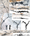
Thin thread-like filaments and filopodia-like tendrils, of varying lengths and thicknesses, are a common feature of macrophages. (A) A single thread-like filament, approximately 120 µm in length (from arrowhead to arrowhead), projects away from the cell body of a macrophage. (B) Multiple tendrils at opposing poles of a macrophage’s soma. (C) Filopodia-like tendrils, located at the tip of the processes of a bipolar macrophage, project away from the cell. Enlarged tracings of the tips of the opposing primary process, highlight the presence of multiple interwoven tendrils. (D) Interwoven, thread-like filaments or filopodia-like tendrils were observed not only along macrophage processes, but also originating from the somata. Images are from whole mounts of the small intestine of adult (A,C,D) and aged (B) rats, demonstrating the presence of these structures regardless of the rat’s age. Macrophages in panels A–D were stained with the antibody to MHCII. Focus stacking was used to create an extended depth of field in panels A–D. Scale bars = 10 µm for all of the panels except for the scale bars in the enlarged tracings of the macrophages in panels C and D where the scale bars = 5 µm.
Figure 5
Figure 5
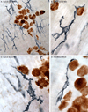
Macrophages are in tight apposition with neurons in the myenteric plexus. (A) Numerous macrophages at the level of the myenteric plexus outline the perimeter of a ganglion. (B–D) The processes of macrophages conform to the surface of the neurons they contact. Images are from whole mounts of the jejunum of aged rats. Whole mounts were double-labeled for HuC/D (neurons; brown) and either CD163 (A–C; blue/grey) or MHCII (D; blue/grey). Focus stacking was used to create an extended depth of field in panel A. Scale bars = 20 µm in A; 10 µm in D (applies to B–D).
Figure 6
Figure 6
Macrophages within the myenteric plexus, viewed at high power (100× oil objective), appeared to make contact with axons and the dendrites of neurons. (A) Axons immunoreactive for nitric oxide synthase (NOS; brown) are in close association with an MHCII+ macrophage (blue/grey). The inset shows the soma of a macrophage in close apposition with a NOS+ axon. (B) Soma and processes of a CD163+ macrophage run parallel to the dendrites of a neuron immunoreactive for the calcium binding protein calbindin (CB; brown). The primary processes of the macrophage in panel B do not make contact with the neuron, but, in the inset, a small tendril originates from the soma of the macrophage and contacts the dendrites of the neuron. (C,E) The processes of two CD163+ macrophages interdigitate with the dendrites of CB+ neurons. (D,F) Enlarged tracings of regions of contact from panels C and E highlight the lock-and-key relationship between macrophages and dendrites. (G,H) An enlargement of two regions from the tracing in panel F. Images are from whole mounts of jejunums from adult (B,E–H) and aged (A,C,D) rats. Focus stacking was used to create an extended depth of field in panels A–C, and E. Scale bars = 10 µm in A–C and E; 5 µm in insets in A,B and D,F; 2.5 µm in G,H.
Figure 7
Figure 7
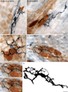
Macrophage contacts with myenteric neurons varied from selective (i.e., individual neurons within a ganglion) to extensive (most or all of the neurons within a ganglion contacted). (A) The pole of a single HuC/D+ neuron (brown) is surrounded by an MHCII+ macrophage (blue/grey). (B) Similar targeting of a neuron immunoreactive for the calcium binding protein calretinin (CR; brown) is carried out by an MHCII+ macrophage. (C) Calretinin-positive (brown) and -negative (unstained) neurons are encircled by multiple processes from a single macrophage with its soma located at the center of the ganglion. (D) An MHCII+ macrophage within a ganglion (five CR+ neurons) with its extensive processes spread throughout the entire ganglion. Several MHCII+ monocytes are in close proximity to the ganglion shown in panel D. (E,F) Different planes of focus of the macrophage in panel D illustrates the soma of the macrophage located on top of the CR+ neurons with its extensively branching processes extending throughout the depth of the ganglion. (G) An enlarged tracing of the macrophage seen in D emphases its size and complexity. Images are from whole mounts of the small intestine of adult (A) and aged (B–D) rats demonstrating that this interaction occurs across the lifespan of the rat. Focus stacking was used to create an extended depth of field in panels B–F. Scale bars = 10 µm; B applies to C; F applies to E.
Figure 8
Figure 8
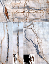
Macrophages contact the varicosities on noradrenergic axons. (A) For comparative purposes, we’ve included an image of a HuC/D+ myenteric neuron (brown) encircled by varicose axons immunoreactive for tyrosine hydroxylase (TH; blue/grey). Varicosities (A; arrowheads) along TH+ axons are thought to be sites of non-synaptic neurotransmitter release. (B) The varicosities of TH+ axons (brown) contact the soma (arrowheads) and process (dotted box) of a CD163+ macrophage (blue/grey). (C–E) Bipolar macrophages immunoreactive for MHCII (blue/grey) are in direct contact with TH+ varicose axons (brown). The inset in panel D is an enlarged tracing of the tip of the macrophage that highlights the two small tendrils (black) along the length of the TH+ axon (brown). The inset in panel E is an enlarged tracing of the point of contact between the MHCII+ macrophage and varicosities on the TH+ axon. Images are from whole mounts of the duodenum (B), jejunum (A,C,D), and ileum (E) adult (C–E), middle-aged (B), and aged (A) rats. Focus stacking was used to create an extended depth of field in panels A–E. Scale bars = 10 µm in A,C–E; 20 µm in B; 1.5 µm for inset in D; 2.5 µm for inset in E.
Figure 9
Figure 9
Macrophages often outlined the perimeter of the myenteric plexus without infiltrating the ganglia (A,B); however, ganglia were observed with varying degrees of infiltration by amoeboid-like macrophages (C–F). Macrophages were immunoreactive for either CD163 (A) or MHCII (B–F). The ganglia were visualized by double labeling for α-SYNC (A, C–F) or TH (B). Images were from the whole mounts of the large intestine (A,B) and stomach (C–F) of adult (A,B), and aged (C–F) rats. Scale bars = 25 µm in B (applies to A,B); 50 µm in F (applies to C–F).
Figure 10
Figure 10
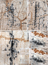
Aggregates or condensations of macrophages, consisting of numerous cells clumped together in large dense masses, occurred in whole mounts stained with the antibody to MHCII (A–E). The loss of morphological definition in an aggregate of MHCII+ macrophages is evident in panel C. The low power (10× objective) inset in panel C illustrates the low density of MHCII+ macrophages in the vicinity of the aggregated macrophages (outlined by the black box). (D,E) Two different focal planes of the smooth muscle wall at the site of an aggregate, illustrate how the core of this particular aggregate of macrophages is located at the level of the deep muscle (D) and not the myenteric plexus (E). The myenteric plexus was identified by double staining for either TH (A,B; brown) or α-SYNC (C; brown). Myenteric neurons were stained with the antibody to HuC/D (D,E; brown). Images are from whole mounts of the jejunum. Focus stacking was used to create an extended depth of field in panels C–E. Scale bars = 50 µm in B (applies to A,B); 25 µm in C (250 µm in inset); 25 µm in E (applies to D,E).
Figure 11
Figure 11
Fragmented macrophages were present within the smooth muscle wall of the GI tract. Fragmented macrophages (dotted outline) occurred in close proximity to neurons (A,B; brown) and axons (D,E; brown), and were immunoreactive for both CD163 (C–E; blue/grey) and MHCII (A,B; blue/grey). Neurons (A,B) were labeled with the antibody to HuC/D while axons were labeled with the antibody to TH (D,E). (C) Macrophages with fragmenting processes and intact soma were observed. The TH+ axons in panels D and E are tangled and swollen suggesting reorganization, degeneration, or both. While macrophages in the tangle of TH+ axons, in the upper left of the panel, still maintain some morphological integrity, small CD163+ fragments are all that remain of macrophages in the tangle of TH+ axons in the bottom right of panel E. Images are from whole mounts of the stomach (D,E) and small intestine (A–C) of middle-aged (D,E) and aged (A–C) rats, ages when plasticity and degeneration of enteric nerves is common. Focus stacking was used to create an extended depth of field in panels A,B,D, and E. Scale bars = 10 µm in C (applies to A–C); 10 µm in D,E.
Figure 12
Figure 12
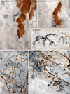
Phagocytosis of aggregated alpha-synuclein (α-SYNC) by macrophages in the GI tract of aged rats. (A) The processes of a CD163+ macrophage (blue/grey) contact a large aggregate of α-SYNC (brown). (B) Small thread-like tendrils originating from a CD163+ macrophage (blue/grey) surround an aggregate of α-SYNC (brown). (C) An enlarged tracing of the tendrils in panel B. Panels A–C are from the stomach of an aged rat. (D) An MHCII+ macrophage (blue/grey) with numerous long-thin tendrils within a field of small α-SYNC+ aggregates (arrowheads) in the jejunum of an adult rat. (E) An enlarged tracing of the macrophage in panel D emphasizing the presence of numerous filopodia and tendrils. Aggregated α-SYNC was rare in the adult enteric nervous system (D), but common in the GI tract of aged (A–C) rats. Focus stacking was used to create an extended depth of field in panels A–C. Scale bars = 10 µm in D (applies to A,B,D); 5 µm in C; 5 µm in E.
Figure 13
Figure 13
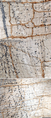
Monocytes and amoeboid-like macrophages (grey/blue) infiltrate the GI musculature and appear, based upon their concentration gradient, to be migrating through the length and width of the small (A,B) and large (C) intestines. Images are from whole mounts stained for MHCII (A,B) and CD163 (C). Whole mounts were double labeled for either α-SYNC (A,B; brown) or TH (C; brown) to visualize the myenteric plexus. Scale bar in C = 125 µm for A,B; 250 µm for C.
Figure 14
Figure 14
The outside perimeter of the blood vessels (BV) located in the smooth muscle wall of the GI tract are well defined by TH+ axons (brown), monocytes, amoeboid-like cells, and macrophages (blue/grey) throughout the GI tract (small intestine: A; large intestine: B–C). Monocytes and macrophages lining the BV are immunoreactive for CD163 (A,C) and MHCII (B). The pattern was the same regardless of age (adult: B,C; middle-aged: A). Panels C is a montage consisting of two low power (20× objective) images. Scale bars = 100 µm in A; 40 µm in B; 50 µm in C.

Similar articles

Cited by

References

    1. Abalo R, Jose Rivera A, Vera G, Isabel Martin M. Ileal myenteric plexus in aged guinea-pigs: loss of structure and calretinin-immunoreactive neurones. Neurogastroenterol Motil. 2005;17(1):123–132. - PubMed
    1. Bauer AJ. Mentation on the immunological modulation of gastrointestinal motility. Neurogastroenterol Motil. 2008;20(Suppl 1):81–90. - PubMed
    1. Bennett MC. The role of alpha-synuclein in neurodegenerative diseases. Pharmacol Ther. 2005;105(3):311–331. - PubMed
    1. Birge RB, Ucker DS. Innate apoptotic immunity: the calming touch of death. Cell death and differentiation. 2008;15(7):1096–1102. - PubMed
    1. Borovikova LV, Ivanova S, Zhang M, Yang H, Botchkina GI, Watkins LR, Wang H, Abumrad N, Eaton JW, Tracey KJ. Vagus nerve stimulation attenuates the systemic inflammatory response to endotoxin. Nature. 2000;405(6785):458–462. - PubMed

Publication types

MeSH terms