. 2012;13(3):R17.
doi: 10.1186/gb-2012-13-3-r17.

The RNA-binding landscapes of two SR proteins reveal unique functions and binding to diverse RNA classes

Affiliations

The RNA-binding landscapes of two SR proteins reveal unique functions and binding to diverse RNA classes

Minna-Liisa Änkö et al. Genome Biol. 2012.

Abstract

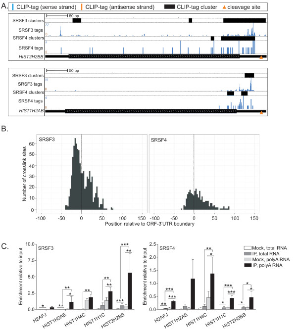
Background: The SR proteins comprise a family of essential, structurally related RNA binding proteins. The complexity of their RNA targets and specificity of RNA recognition in vivo is not well understood. Here we use iCLIP to globally analyze and compare the RNA binding properties of two SR proteins, SRSF3 and SRSF4, in murine cells.

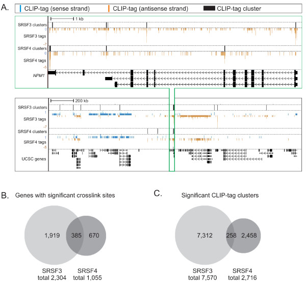
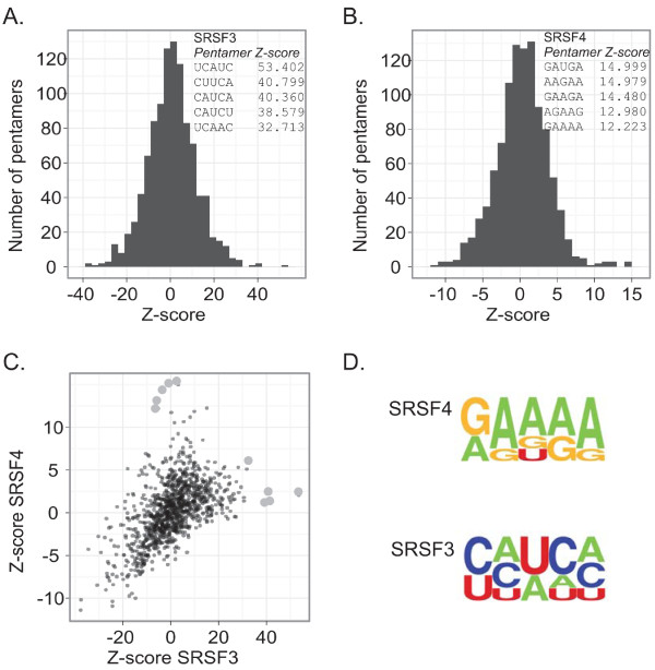
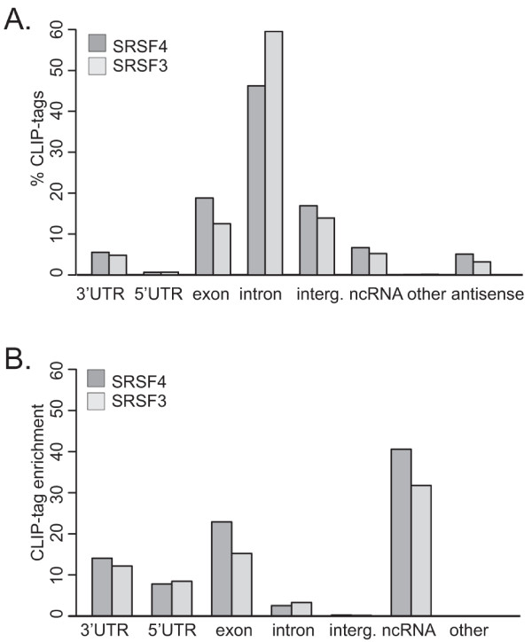
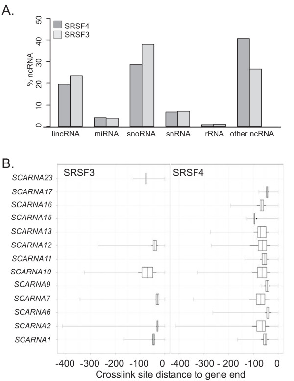
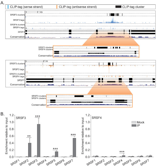
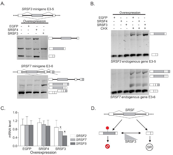
Results: SRSF3 and SRSF4 binding sites mapped to largely non-overlapping target genes, and in vivo consensus binding motifs were distinct. Interactions with intronless and intron-containing mRNAs as well as non-coding RNAs were detected. Surprisingly, both SR proteins bound to the 3' ends of the majority of intronless histone transcripts, implicating SRSF3 and SRSF4 in histone mRNA metabolism. In contrast, SRSF3 but not SRSF4 specifically bound transcripts encoding numerous RNA binding proteins. Remarkably, SRSF3 was shown to modulate alternative splicing of its own as well as three other transcripts encoding SR proteins. These SRSF3-mediated splicing events led to downregulation of heterologous SR proteins via nonsense-mediated decay.

Conclusions: SRSF3 and SRSF4 display unique RNA binding properties underlying diverse cellular regulatory mechanisms, with shared as well as unique coding and non-coding targets. Importantly, CLIP analysis led to the discovery that SRSF3 cross-regulates the expression of other SR protein family members.


Figures

Figure 1
Figure 1
SRSF3 and SRSF4 CLIP-tags cluster to distinct positions in mouse RNAs. (a) NPM1 gene (green box) and the surrounding approximately 3 MB region in chromosome 11 (black box) with SRSF3 and SRSF4 CLIP-tags and clusters. The numbers on the left represent the number of CLIP-tags within the window. The sense strand is marked in blue and the antisense strand in orange. Note that the genes in the antisense strand run from right to left. (b) Comparison of annotated genes with significant SRSF3 or SRSF4 crosslink sites (false discovery rate < 0.05). (c) Comparison of significant SRSF3 and SRSF4 CLIP-tag clusters (overlap of clusters ≥ 15 nucleotides).
Figure 2
Figure 2
In vivo binding specificity of SRSF3 and SRSF4. (a, b) The frequency distribution of SRSF3 (a) and SRSF4 (b) pentamer Z-scores. The Z-score was calculated relative to randomized genomic positions by shuffling the crosslink positions 100 times within the genes. Five pentamers with highest Z-scores are shown. (c) Correlation of SRSF3 and SRSF4 pentamer Z-scores. The top five pentamers presented in (a, b) are marked as larger light grey dots. (d) Consensus motifs were derived from the top pentamers shown in (a, b).
Figure 3
Figure 3
Distribution of SRSF3 and SRSF4 CLIP-tags within RNA classes and transcript regions. (a) The proportion of CLIP-tags that mapped to different RNAs relative to the total number of CLIP-tags. (b) The fold enrichment of CLIP-tag density (the number of CLIP-tags divided by the length of each RNA feature) in different RNAs relative to the average CLIP-tag density in the genome.
Figure 4
Figure 4
ncRNAs with SRSF3 and SRSF4 crosslink sites. (a) The distribution of crosslink sites within the ncRNA subclasses. (b) The position of the SRSF4 CLIP-tag clusters relative to the scaRNA 3' end. 'Other ncRNAs' are processed transcripts with no known ORF or function.
Figure 5
Figure 5
SRSF3 and SRSF4 bind to numerous intronless histone mRNAs at a consistent position. (a) SRSF3 and SRSF4 CLIP-tags and clusters in HIST2H2BB and HIST1H2AB genes. Labels as in Figure 1a. The orange arrowheads mark the mRNA 3' end cleavage site. (b) Mapping of SRSF3 (left panel) and SRSF4 (right panel) crosslink sites to the ORF-3' UTR boundary of histone mRNAs. The position 0 marked with a dotted line represents the ORF-3' UTR boundary. (c) Cytoplasmic levels of histone mRNAs associated with SRSF3 or SRSF4 determined by UV-RNA immunoprecipitation and reverse transcription quantitative PCR. To prime the reverse transcription reactions, hexamers were used to detect total and oligo-dT to detect polyadenylated histone mRNAs. Data are presented relative to the input sample. Mock is the non-immune control. *P < 0.05, **P < 0.01, ***P < 0.001 (Student's unpaired t-test, n = 3-6). Error bars are standard deviation. IP, immunoprecipitation.
Figure 6
Figure 6
SRSF3 and SRSF4 contact exons and introns. (a) SRSF3 and SRSF4 crosslink sites mapped around the 5' and 3' splice sites. The position 0 (dotted line) represents the indicated 5' or 3' splice site; the y-axis represents normalized crosslink sites per 103 nucleotides. The normalization is based on the length distribution of exons and introns (Figure S5 in Additional file 1). The data were smoothed using a Gaussian window (half-width of the window = 5). (b) SRSF3 and SRSF4 crosslink sites mapped to predicted mouse branch points. The position 0 (dotted line) represents the branch point nucleotide. Smoothing as in (a).
Figure 7
Figure 7
SRSF3 binds to poison cassette exons in SR proteins. (a) SRSF3 and SRSF4 CLIP-tags and clusters around the alternative cassette exon of SRSF3 and SRSF7 genes. Labels as in Figure 1a. The zoom in represents the ultraconserved regions identified in [42,43]. Note that the genes in the antisense strand run from right to left. (b) The enrichment of mRNAs encoding different SR protein family members after UV crosslinking and SRSF3 or SRSF4 immunoprecipitation (IP). To prime the RT reactions, hexamers were used. Data are presented relative to the input sample. IP is the specific immunoprecipitation and mock is the non-immune control. *P < 0.05, **P < 0.01, ***P < 0.001 (Student's unpaired t-test, n = 3-6). Error bars are standard deviation.
Figure 8
Figure 8
SRSF3 controls the level of SR proteins through splicing regulation. (a) The splicing products of SRSF3 and SRSF7 minigenes determined after 24-hour over-expression of SRSF3, SRSF4 or EGFP (control). The alternative exons are marked with light grey. (b) The splicing products of endogenous SRSF3 and SRSF7 after inhibition of NMD by a 3-hour treatment with cycloheximide (CHX). (c) The expression level of endogenous, mature SRSF2, SRSF5 and SRSF7 mRNAs upon EGFP, SRSF3 or SRSF4 overexpression (24 hours) as measured by RT-qPCR. *P < 0.05 (one-way ANOVA). Error bars are standard deviation. ACTB was used as the reference gene. (d) Schematic showing how SRSF3 controls the levels of other SR protein family members through alternative splicing. The inclusion of a poison cassette exon harboring a premature termination codon (PTC, red stop sign) leads to RNA degradation through NMD.

References

    1. Letunic I, Doerks T, Bork P. SMART 6: recent updates and new developments. Nucleic Acids Res. 2009;37:D229–D232. doi: 10.1093/nar/gkn808. - DOI - PMC - PubMed
    1. Zhong X-Y, Wang P, Han J, Rosenfeld MG, Fu X-D. SR proteins in vertical integration of gene expression from transcription to RNA processing to translation. Mol Cell. 2009;35:1–10. doi: 10.1016/j.molcel.2009.06.016. - DOI - PMC - PubMed
    1. Shepard P, Hertel K. The SR protein family. Genome Biol. 2009;10:242. doi: 10.1186/gb-2009-10-10-242. - DOI - PMC - PubMed
    1. Lin S, Fu XD. SR proteins and related factors in alternative splicing. Adv Exp Med Biol. 2007;623:107–122. doi: 10.1007/978-0-387-77374-2_7. - DOI - PubMed
    1. Sapra AK, Änkö M-L, Grishina I, Lorenz M, Pabis M, Poser I, Rollins J, Weiland E-M, Neugebauer KM. SR protein family members display diverse activities in the formation of nascent and mature mRNPs in vivo. Mol Cell. 2009;34:179–190. doi: 10.1016/j.molcel.2009.02.031. - DOI - PubMed

Publication types